. 2016 May 31;113(22):6289-94.
doi: 10.1073/pnas.1600638113. Epub 2016 May 17.

Incoming human papillomavirus type 16 genome resides in a vesicular compartment throughout mitosis

Affiliations

Incoming human papillomavirus type 16 genome resides in a vesicular compartment throughout mitosis

Stephen DiGiuseppe et al. Proc Natl Acad Sci U S A. .

Abstract

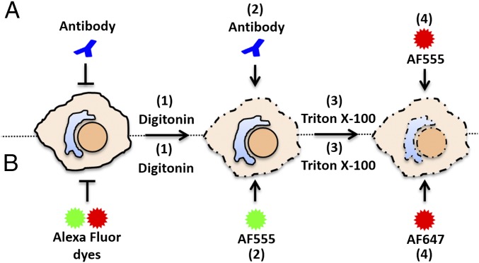
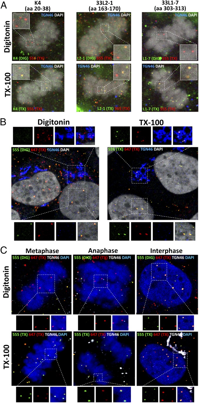
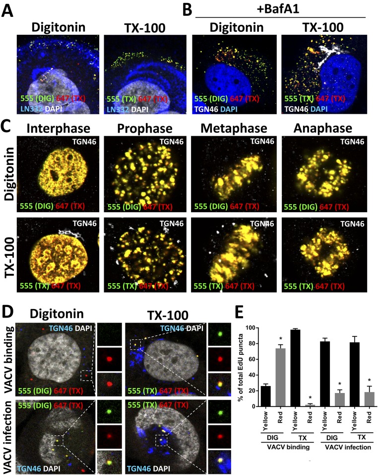
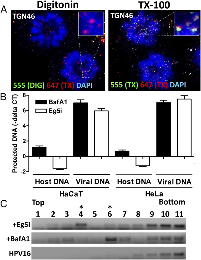
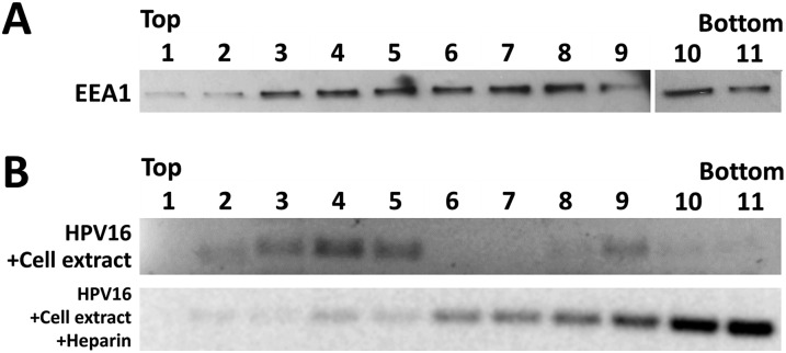
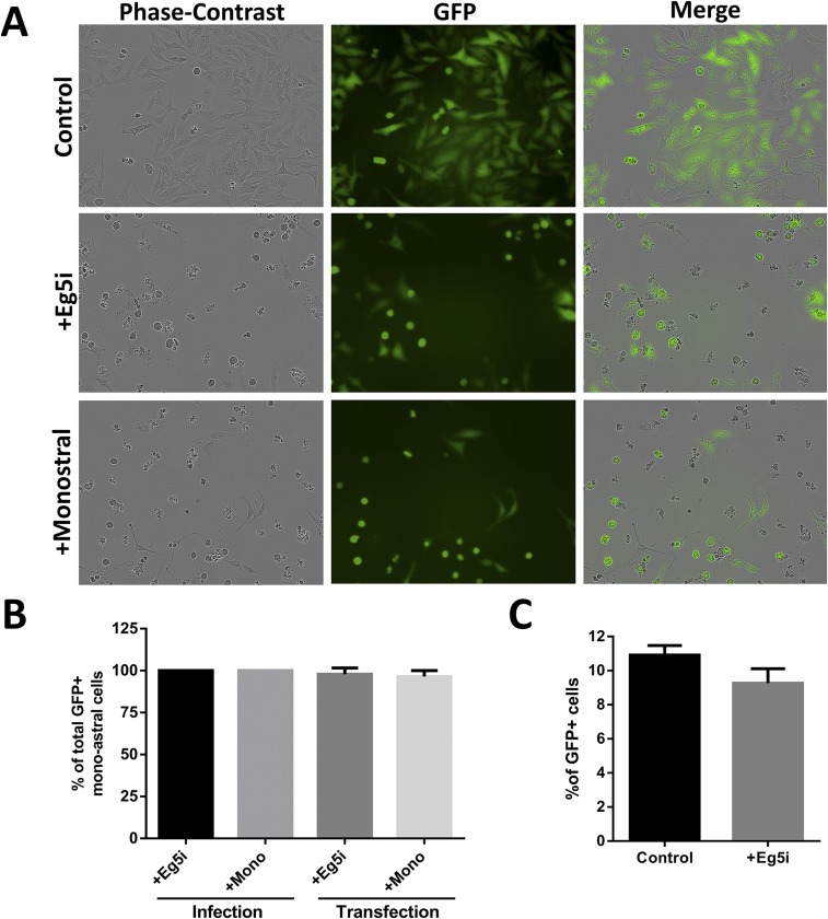
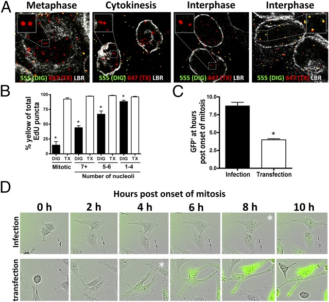
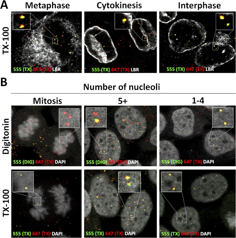
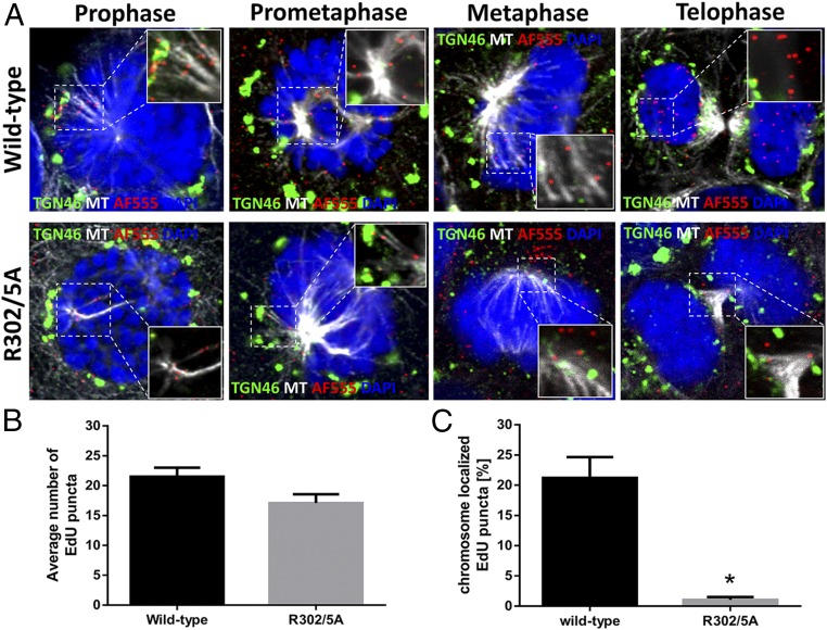
During the entry process, the human papillomavirus (HPV) capsid is trafficked to the trans-Golgi network (TGN), whereupon it enters the nucleus during mitosis. We previously demonstrated that the minor capsid protein L2 assumes a transmembranous conformation in the TGN. Here we provide evidence that the incoming viral genome dissociates from the TGN and associates with microtubules after the onset of mitosis. Deposition onto mitotic chromosomes is L2-mediated. Using differential staining of an incoming viral genome by small molecular dyes in selectively permeabilized cells, nuclease protection, and flotation assays, we found that HPV resides in a membrane-bound vesicle until mitosis is completed and the nuclear envelope has reformed. As a result, expression of the incoming viral genome is delayed. Taken together, these data provide evidence that HPV has evolved a unique strategy for delivering the viral genome to the nucleus of dividing cells. Furthermore, it is unlikely that nuclear vesicles are unique to HPV, and thus we may have uncovered a hitherto unrecognized cellular pathway that may be of interest for future cell biological studies.

Keywords: HPV entry; digitonin; mitosis; nuclear vesicle; vesicular transport.


Conflict of interest statement

The authors declare no conflict of interest.

Figures

Fig. 1.
Fig. 1.
Diagram of differential staining assay for antibodies or Alexa Fluor dyes. (A) (1) Cells infected with EdU-labeled pseudovirus are fixed and permeabilized with DIG or TX-100, (2) incubated with primary and secondary antibodies and fixed briefly again, (3) permeabilized with TX-100, and (4) treated with AF555 (red) in Click-iT reaction buffer. (B) (1) Infected cells are fixed, permeabilized with DIG or TX-100, (2) treated with AF555 (green) in Click-iT reaction buffer, (3) permeabilized with TX-100, and (4) treated with AF647 (red) in Click-iT reaction buffer.
Fig. 2.
Fig. 2.
Incoming viral genome resides in a vesicle until the completion of mitosis. (A) At 24 hpi with EdU-labeled pseudovirus, HaCaT cells were fixed, permeabilized with 5 µg/mL of DIG or 0.5% TX-100, and incubated with primary L2-specific antibodies K4 (amino acids 20–38), 33L2-1 (amino acids 163–170), or L1-specific antibody 33L1-7 (amino acids 303–313), followed by a subsequent secondary antibody (green). Cells were fixed again, permeabilized with 0.5% TX-100, and treated with AF555 (red) in Click-iT reaction buffer. The nucleus was stained with DAPI (white). A luminal epitope of TGN46 (blue) served as a control to assess intracellular membrane integrity. Note the lack of reactivity of both the K4 and 33L1-7 antibodies in DIG-treated cells. (B) HaCaT cells were infected as described above. At 24 hpi, cells were fixed, permeabilized in 0.625 µg/mL DIG or 0.5% TX-100, and treated with AF555 (green) in Click-iT reaction buffer. Cells were fixed again, permeabilized with 0.5% TX-100, and treated with AF647 (red). TGN46 (blue) was stained after TX-100 treatment. The nucleus was stained with DAPI (white). (C) HaCaT cells were infected and differentially stained as described in B. A luminal epitope of TGN46 (white) served as a control to assess intracellular membrane integrity. Note the absence of AF555 staining in pseudogenomes localized to the TGN and associated with mitotic chromosomes.
Fig. S1.
Fig. S1.
Differential staining controls. (A) At 1 hpi, HaCaT cells were fixed, permeabilized with 0.625 µg/mL DIG or 0.5% TX-100, and treated with AF555 (green) in Click-iT reaction buffer. Cells were briefly fixed again, permeabilized with 0.5% TX-100, and treated with AF647 (red) in Click-iT reaction buffer. The ECM marker Laminin 332 (LN332) (blue) was stained after TX-100 treatment. The nuclei are stained with DAPI (white). Note that the EdU puncta localized on the ECM is accessible to AF555 after DIG treatment. (B) HaCaT cells were infected with EdU-labeled HPV16 pseudovirus for 18 h in the presence of 1 µM BafA1. Differential staining of pseudogenomes was performed as described in A. A luminal epitope of TGN46 (white) served as a control to assess intracellular membrane integrity. (C) Uninfected HaCaT cells were pulse-labeled with 10 µM EdU for 4 h, then differentially stained as described in A. A luminal epitope of TGN46 (white) served as a control to assess intracellular membrane integrity. Note that host chromatin was accessible to AF555 after DIG treatment in interphase and all phases of mitosis. (D) HaCaT cells were infected with VACV for 1 h at 4 °C to allow binding. Cells were either directly fixed or shifted to 37 °C for 2 h and then fixed. Differential staining of VACV genomes was performed as described in A. A luminal epitope of TGN46 (white) served as a control to assess intracellular membrane integrity. (E) Quantification of dye accessibility to the VACV genome at 1 h after binding or 2 h after entry. Quantification is based on the number of double-positive EdU puncta as a percentage of total EdU puncta per image (n = 15 images). All P values are < 0.0005. Note that VACV genome localized at the cell surface was inaccessible to AF555, but became accessible after internalization in DIG-treated cells.
Fig. 3.
Fig. 3.
Vesicle protects the incoming viral genome from digestion during mitosis. (A) HaCaT cells were infected in the presence of Eg5i and differentially stained as described in Fig. 2 B and C. Note the absence of AF555 staining in pseudogenomes associated with arrested cells. (B) HaCaT and HeLa cells were infected with HPV16 pseudovirus for 24 h and 18 h, respectively, in the presence of 1.5 µM Eg5i or 0.5 µM BafA1. Monoastral phenotypic cells were isolated from the bulk population of Eg5i-treated cells, whereas total cells were collected in the BafA1 treatment. Note that BafA1 treatment blocks acidification and capsid uncoating. Plasma membranes were mechanically disrupted, and the cell lysates were treated with 10 U of DNase I for 3 h in the presence or absence of TX-100. Total DNA was isolated and analyzed by real-time quantitative PCR (qPCR). Data are reported as –Δ Ct normalized to DNA isolated from TX-100–treated samples. Note that the HPV16 genome is protected from DNase I digestion up to 6.99 ± 0.23 and 7.02 ± 0.18 Ct values in HaCaT and HeLa cells, respectively (n = 3). (C) HeLa cells were infected with HPV16 quasivirions encapsidating an HPV16 genome for 24 h in the presence of 1.5 µM Eg5i or 1 µM BafA1. Cells were collected and mechanically disrupted as described in B. Cell lysates were fractionated using a sucrose density gradient. DNA in the gradient fractions was amplified using HPV16 E6-specific primers by conventional PCR using dilutions of the gradient fractions as a template and visualized by gel electrophoresis. Purified HPV16 quasivirus was fractionated as a virus-only control. Note that the HPV16 genome is enriched in fraction 4 in the Eg5i-treated cells, whereas it is enriched in fraction 6 in BafA1-treated cells.
Fig. S2.
Fig. S2.
Separation of vesicles via the sucrose flotation assay. (A) Uninfected HeLa cells were grown for 18 h, trypsinized, and then collected. Cellular homogenates were prepared by incubating cells on ice for 15 min in SFB (20 mM Hepes pH 7.4, 10 mM KCl, 1.5 mM MgCl2, and 1 mM EDTA) and then passing the cells 20 times through a 27-gauge needle. The PNS was adjusted to 40.6% sucrose in 1 mL total volume, and then placed into the bottom of an SW55 centrifuge tube. The sucrose flotation assay column was generated by overlaying the PNS with 2.5 mL of 35% sucrose, 1 mL of 25% sucrose, and 0.5 mL of 8.56% sucrose solution in SFB. Fractions of ∼425 µL were collected from the top after the column was spun down using a SW55 rotor for 40,000 rpm at 5 °C. The fractions were collected and probed for the presence of EEA1 by immunoblotting. Note that EEA1 is found in fractions near the top of the gradient. (B) Highly purified virus stock was combined with cell extract and subjected to the sucrose flotation assay as described in A. As a control, highly purified virus stock was preincubated with 100 µg/mL of heparin at 37 °C for 1 h, combined with cell extract, and subjected to sucrose flotation assay as described in A. Fractions were collected and used as a template for conventional PCR using E6-specific primers to detect the presence of the viral DNA. Note that preincubation with heparin blocked upward migration of the viral DNA.
Fig. S3.
Fig. S3.
Arrest in mitosis with inhibitors of Eg5 is leaky. (A) HeLa cells were infected with HPV16 pseudovirus encapsidating a GFP expression plasmid pseudogenome for 72 h in the presence or absence of 1.5 µM Eg5i and 100 µM monastrol. Cells were monitored by live-cell imaging. Images from 48 h are presented here. Note that GFP expression is visible in cells that exhibit the monoastral phenotype. Also note that there are markedly less viable cells after 48 h in the Eg5-inhibited cells. (B) Quantification for the number of GFP+ monoastral phenotypic cells that have undergone at least one round of division before expressing GFP (∼30 cells counted per biological replicate; n = 3) as observed by live-cell imaging. Note that ∼100% of cells that enter the monoastral phenotype have undergone at least one round of mitosis before expressing GFP. (C) HeLa cells were infected with HPV16 pseudovirus for 48 h in the presence or absence of 1.5 µM Eg5i, and the number of GFP+ cells were counted by flow cytometry. Note that Eg5i treatment did not show any loss in infectivity as measured by FACS.
Fig. 4.
Fig. 4.
Release of the viral genome from the vesicle is delayed. (A) HaCaT cells were infected as described in Fig. 2 B and C. LBR (white) was stained after TX-100 treatment. Note the absence of AF555 staining within the nuclei of the DIG-treated cells in cytokinesis and some interphase cells. (B) Quantification of the AF555 accessibility as a percentage of total AF647 relative to the number of individual puncta per cell. The total number of nucleoli per cell was counted as well (∼50 cells counted per biological replicate, n = 3; mitotic, P < 0.0005; 7+ nucleoli, P < 0.0001; 5–6 nucleoli, P < 0.005; 1–4 nucleoli, P = 0.0303). Note that increased accessibility is inversely correlated with the number of nucleoli present per image slice. (C and D) HeLa cells were infected with HPV16 pseudovirus encapsidating pseudogenomes or transfected using MATra with pfwB plasmid for 72 h. Live-cell images were obtained once an hour. Cells were quantified by counting how long it took cells to display GFP expression following the onset of mitosis (50 cells counted per biological replicate, n = 3; P < 0.005). (D) Time lapse of HeLa cells from the onset of mitosis until display of GFP expression after delivery of pfwB by HPV16 pseudovirus and transfection with MATra reagent, respectively. Expression levels of GFP in the infection reached comparable levels to those of the transfection over time (Movie S3).
Fig. S4.
Fig. S4.
Pseudogenome becomes accessible during early interphase. (A) HaCaT cells were infected with EdU-labeled HPV16 pseudovirus for 24 h. Cells were fixed, permeabilized with 0.5% TX-100, and treated with AF555 (green) in Click-iT reaction buffer. Cells were fixed again, permeabilized with 0.5% TX-100, and treated with AF647 (red) in Click-iT reaction buffer. LBR (white) was stained. Note that the pseudogenome was accessible to AF555 during cytokinesis and early interphase after TX-100 permeabilization. (B) HaCaT cells were infected with EdU-labeled HPV16 pseudovirus for 24 h and stained. Cells were fixed, permeabilized with 0.625 µg/mL DIG, and treated with AF555 (green) in Click-iT reaction buffer. Then the cells were fixed again, permeabilized with 0.5% TX-100, and treated with AF647 (red) in Click-iT reaction buffer. The nuclei were stained with DAPI (white). Note that the pseudogenomes exhibited limited accessibility to AF555 after DIG treatment in cells with five or more nucleoli.
Fig. 5.
Fig. 5.
The incoming viral genome associates with microtubules during mitosis, and L2 facilitates delivery to the nucleus. (A) HaCaT cells were infected for 24 h with pseudovirus harboring either WT 16L2 or 16L2-R302/5A that encapsidated an EdU-labeled pseudogenome. Cells were fixed, permeabilized in 0.5% TX-100, and treated with AF555 in Click-iT reaction buffer to detect the pseudogenomes (red). The TGN (green) or microtubules (MT; white) were stained as well. The nuclei are stained with DAPI (blue). Still images of cells in different stages of mitosis were collected. Note that the pseudogenome dissociated from TGN46 marker during prophase and associated with microtubules throughout mitosis. (B) Graphical representation of the average number of EdU puncta in three continuous Z-stack images per cell counted (n = 15 cells; P = 0.0431). (C) Graphical representation for the percentage of EdU puncta localized to condensed chromosomes of cells in different stages of mitosis for both the WT and mutant pseudoviruses. Quantification is based on the percentage of EdU puncta localized to condensed chromosomes out of the total number of EdU puncta per cell based on three continuous Z-stack images per cell counted (n = 15 cells; P < 0.0001).

Similar articles

Cited by

References

    1. Paludan SR, Bowie AG. Immune sensing of DNA. Immunity. 2013;38(5):870–880. - PMC - PubMed
    1. Bienkowska-Haba M, Williams C, Kim SM, Garcea RL, Sapp M. Cyclophilins facilitate dissociation of the human papillomavirus type 16 capsid protein L1 from the L2/DNA complex following virus entry. J Virol. 2012;86(18):9875–9887. - PMC - PubMed
    1. Buck CB, et al. Arrangement of L2 within the papillomavirus capsid. J Virol. 2008;82(11):5190–5197. - PMC - PubMed
    1. Buck CB, Pastrana DV, Lowy DR, Schiller JT. Generation of HPV pseudovirions using transfection and their use in neutralization assays. Methods Mol Med. 2005;119:445–462. - PubMed
    1. Modis Y, Trus BL, Harrison SC. Atomic model of the papillomavirus capsid. EMBO J. 2002;21(18):4754–4762. - PMC - PubMed

Publication types

MeSH terms