Review
. 2016 Sep 7;37(34):2645-57.
doi: 10.1093/eurheartj/ehw096. Epub 2016 Mar 31.

Low-gradient aortic stenosis

Affiliations
Review

Low-gradient aortic stenosis

Marie-Annick Clavel et al. Eur Heart J. .

Abstract

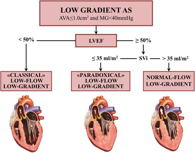
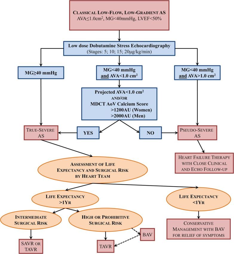
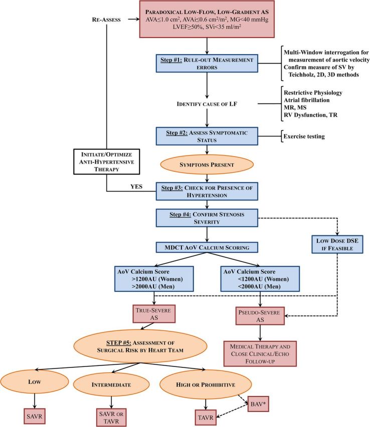
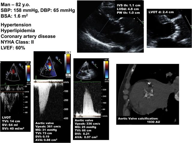
An important proportion of patients with aortic stenosis (AS) have a 'low-gradient' AS, i.e. a small aortic valve area (AVA <1.0 cm(2)) consistent with severe AS but a low mean transvalvular gradient (<40 mmHg) consistent with non-severe AS. The management of this subset of patients is particularly challenging because the AVA-gradient discrepancy raises uncertainty about the actual stenosis severity and thus about the indication for aortic valve replacement (AVR) if the patient has symptoms and/or left ventricular (LV) systolic dysfunction. The most frequent cause of low-gradient (LG) AS is the presence of a low LV outflow state, which may occur with reduced left ventricular ejection fraction (LVEF), i.e. classical low-flow, low-gradient (LF-LG), or preserved LVEF, i.e. paradoxical LF-LG. Furthermore, a substantial proportion of patients with AS may have a normal-flow, low-gradient (NF-LG) AS: i.e. a small AVA-low-gradient combination but with a normal flow. One of the most important clinical challenges in these three categories of patients with LG AS (classical LF-LG, paradoxical LF-LG, and NF-LG) is to differentiate a true-severe AS that generally benefits from AVR vs. a pseudo-severe AS that should be managed conservatively. A low-dose dobutamine stress echocardiography may be used for this purpose in patients with classical LF-LG AS, whereas aortic valve calcium scoring by multi-detector computed tomography is the preferred modality in those with paradoxical LF-LG or NF-LG AS. Although patients with LF-LG severe AS have worse outcomes than those with high-gradient AS following AVR, they nonetheless display an important survival benefit with this intervention. Some studies suggest that transcatheter AVR may be superior to surgical AVR in patients with LF-LG AS.

Keywords: Aortic stenosis; Aortic valve replacement; Computed tomography; Echocardiography; Low flow; Low gradient; Transcatheter aortic valve replacement.


Figures

Figure 1
Figure 1
Subtypes of low-gradient aortic stenosis. AS, aortic stenosis; AVA, aortic valve area; LVEF, left ventricular ejection fraction; MG, mean transvalvular gradient; SVi, stroke volume index.
Figure 2
Figure 2
Algorithm for the management of classical (reduced left ventricular ejection fraction) low-flow, low-gradient aortic stenosis. AoV, aortic valve; BAV, balloon aortic valvuloplasty; MDCT, multi-detector computed tomography; SAVR, surgical aortic valve replacement; TAVR, transcatheter aortic valve replacement. Other abbreviations as in Figure 1.
Figure 3
Figure 3
Dobutamine stress echocardiography for the assessment of stenosis severity in classical (reduced left ventricular ejection fraction), low-flow, low-gradient aortic stenosis. (A) Patient with true-severe aortic stenosis; (B) patient with pseudo-severe aortic stenosis; (C and D) patient with persisting discordant grading (i.e. small aortic valve area with low mean transvalvular gradient) at dobutamine stress echocardiography (C) and calculation of the projected aortic valve area at normal flow rate (0.84 cm2) to confirm the presence of severe stenosis in this patient (D). (E and F) Patient with no left ventricular flow reserve on dobutamine stress echocardiography and measurement of aortic valve calcification score by multi-detector computed tomography to confirm stenosis severity. This patient is a woman and has a calcium score of 1802 AU, which is consistent with true-severe aortic stenosis (see Figure 4). DSE, dobutamine stress echocardiography; LVOT, LV outflow tract; LVOTd, LVOT diameter; TVI, time-velocity integral; SV, stroke volume; SVRest, SV at rest; SVPeak, SV at peak DSE; Q, mean transvalvular flow rate (SV/ LV ejection time); VPeak, peak aortic jet velocity. Other abbreviations as in Figures 1 and 2.
Figure 3
Figure 3
Dobutamine stress echocardiography for the assessment of stenosis severity in classical (reduced left ventricular ejection fraction), low-flow, low-gradient aortic stenosis. (A) Patient with true-severe aortic stenosis; (B) patient with pseudo-severe aortic stenosis; (C and D) patient with persisting discordant grading (i.e. small aortic valve area with low mean transvalvular gradient) at dobutamine stress echocardiography (C) and calculation of the projected aortic valve area at normal flow rate (0.84 cm2) to confirm the presence of severe stenosis in this patient (D). (E and F) Patient with no left ventricular flow reserve on dobutamine stress echocardiography and measurement of aortic valve calcification score by multi-detector computed tomography to confirm stenosis severity. This patient is a woman and has a calcium score of 1802 AU, which is consistent with true-severe aortic stenosis (see Figure 4). DSE, dobutamine stress echocardiography; LVOT, LV outflow tract; LVOTd, LVOT diameter; TVI, time-velocity integral; SV, stroke volume; SVRest, SV at rest; SVPeak, SV at peak DSE; Q, mean transvalvular flow rate (SV/ LV ejection time); VPeak, peak aortic jet velocity. Other abbreviations as in Figures 1 and 2.
Figure 4
Figure 4
Quantitation of aortic valve calcium by multi-detector computed tomography for the assessment of stenosis severity in low-gradient aortic stenosis. (A) Multi-detector computed tomography can be used to quantitate aortic valve calcification by the modified Agatston method. With this method, calcification is defined as four adjacent pixels with density >130 Hounsfield units. Different cut-point values of valve calcium score should be used in women (>1200 AU) vs. men (>2000 AU) to differentiate true-severe vs. pseudo-severe stenosis in low-flow, low-gradient aortic stenosis. (B) Serial multi-detector computed tomography slices at the level of the aortic valve showing a severely calcified valve with a calcium score of 5040 AU consistent with true-severe aortic stenosis. Calcified areas are displayed in yellow in the bottom images. (C) Mild calcification (score 271 AU) consistent with pseudo-severe aortic stenosis. (D) Pitfalls in the assessment of aortic valve calcification by multi-detector computed tomography. For the calculation of calcium score, it is important to only include aortic valve calcification and exclude calcification of aorta, coronary arteries, LVOT, and mitral annulus. Abbreviations as in Figures 1 and 2.
Figure 4
Figure 4
Quantitation of aortic valve calcium by multi-detector computed tomography for the assessment of stenosis severity in low-gradient aortic stenosis. (A) Multi-detector computed tomography can be used to quantitate aortic valve calcification by the modified Agatston method. With this method, calcification is defined as four adjacent pixels with density >130 Hounsfield units. Different cut-point values of valve calcium score should be used in women (>1200 AU) vs. men (>2000 AU) to differentiate true-severe vs. pseudo-severe stenosis in low-flow, low-gradient aortic stenosis. (B) Serial multi-detector computed tomography slices at the level of the aortic valve showing a severely calcified valve with a calcium score of 5040 AU consistent with true-severe aortic stenosis. Calcified areas are displayed in yellow in the bottom images. (C) Mild calcification (score 271 AU) consistent with pseudo-severe aortic stenosis. (D) Pitfalls in the assessment of aortic valve calcification by multi-detector computed tomography. For the calculation of calcium score, it is important to only include aortic valve calcification and exclude calcification of aorta, coronary arteries, LVOT, and mitral annulus. Abbreviations as in Figures 1 and 2.
Figure 5
Figure 5
Patient with paradoxical (preserved left ventricular ejection fraction) low-flow, low-gradient severe aortic stenosis. This case underlines the importance of multi-window continuous-wave Doppler interrogation for the measurement of the aortic velocity and gradient. In this patient, the gradient was higher at the right parasternal window than at the apical window. It is also important to rule-out error in the measurement of stroke volume. In this patient, the stroke volume measured by pulsed wave Doppler in the LVOT (48 mL) is corroborated by the stroke volume obtained with the modified Teichholz method: LV end-diastolic volume by Teichholz (70 mL) × left ventricular ejection fraction by biplane Simpson (70%) = 49 mL. The patient is in low flow (SVi: 28 mL/m2). Aortic valve calcium score by multi-detector computed tomography corroborates presence of true-severe stenosis. ACEI, angiotensin conversion enzyme inhibitor; BSA, body surface area; DVI, Doppler velocity index; LVDd, LV end-diastolic diameter; IVS th, interventricular septum thickness; PWth, posterior wall thickness; SBP/DBP, systolic and diastolic blood pressure. Other abbreviations as in Figures 1, 2, and 3.
Figure 6
Figure 6
Algorithm for the management of paradoxical (preserved left ventricular ejection fraction) low-flow, low-gradient aortic stenosis. AVAi, indexed aortic valve area; MR, mitral regurgitation; MS, mitral stenosis; TR, tricuspid regurgitation; other abbreviations as in Figures 1, 2, and 3.
Figure 7
Figure 7
Patient with normal-flow, low-gradient aortic stenosis. This patient has a normal flow (stroke volume index >35 mL/m2), small aortic valve area, small indexed aortic valve area, and low mean transvalvular gradient at echocardiography. As in the patient with paradoxical low-flow, low-gradient (Figure 5), it is important first to rule-out measurement errors and then confirm stenosis severity, especially if the patient is symptomatic. In this patient, aortic valve calcium score by multi-detector computed tomography suggests the presence of a moderate to severe aortic stenosis.

Comment in

Similar articles

Cited by

References

    1. Vahanian A, Alfieri O, Andreotti F, Antunes MJ, Baron-Esquivias G, Baumgartner H, Borger MA, Carrel TP, De BM, Evangelista A, Falk V, Iung B, Lancellotti P, Pierard L, Price S, Schafers HJ, Schuler G, Stepinska J, Swedberg K, Takkenberg J, von Oppell UO, Windecker S, Zamorano JL, Zembala M. Guidelines on the management of valvular heart disease (version 2012). Joint task force on the management of valvular heart disease of the European Society of Cardiology (ESC); European Association for Cardio-Thoracic Surgery (EACTS). Eur Heart J 2012;33:2451–2496. - PubMed
    1. Nishimura RA, Otto CM, Bonow RO, Carabello BA, Erwin JP, Guyton RA, O'Gara PT, Ruiz CE, Skubas NJ, Sorajja P, Sundt TM, Thomas JD. 2014 AHA/ACC guideline for the management of patients with valvular heart disease: executive summary. A report of the American College of Cardiology/American heart association task force on practice guidelines. J Am Coll Cardiol 2014;63:2438–2488. - PubMed
    1. Hachicha Z, Dumesnil JG, Bogaty P, Pibarot P. Paradoxical low flow, low gradient severe aortic stenosis despite preserved ejection fraction is associated with higher afterload and reduced survival. Circulation 2007;115:2856–2864. - PubMed
    1. Minners J, Allgeier M, Gohlke-Baerwolf C, Kienzle RP, Neumann FJ, Jander N. Inconsistent grading of aortic valve stenosis by current guidelines: haemodynamic studies in patients with apparently normal left ventricular function. Heart 2010;96:1463–1468. - PubMed
    1. Lancellotti P, Magne J, Donal E, Davin L, O'Connor K, Rosca M, Szymanski C, Cosyns B, Piérard LA. Clinical outcome in asymptomatic severe aortic stenosis. Insights from the new proposed aortic stenosis grading classification. J Am Coll Cardiol 2012;59:235–243. - PubMed