

Original Research: Influence of okadaic acid on hyperphosphorylation of tau and nicotinic acetylcholine receptors in primary neurons
- PMID: 27190248
- PMCID: PMC5027944
- DOI: 10.1177/1535370216650759
Original Research: Influence of okadaic acid on hyperphosphorylation of tau and nicotinic acetylcholine receptors in primary neurons
Abstract
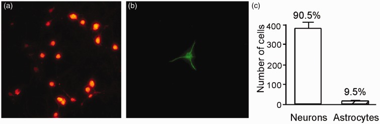
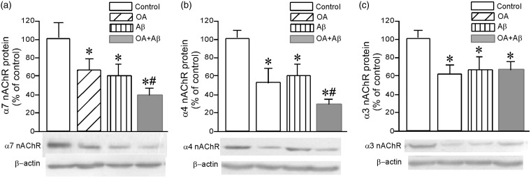
The aim of the study was to investigate the influence of hyperphosphorylation of tau induced by okadaic acid on the expression of nicotinic acetylcholine receptors and the neurotoxicity of β-amyloid peptide. Primary cultures of neurons isolated from the hippocampus of the brains of neonatal rats were exposed to okadaic acid or/and Aβ1-42 Tau phosphorylated at Ser404 and Ser202, and the protein expressions of α7, α4 and α3 nAChR subunits were quantified by Western blotting, and their corresponding mRNAs by real-time PCR. Superoxide dismutase activity was assayed biochemically and malondialdehyde by thiobarbituric acid-reactive substance. As compared to controls, phosphorylations of tau at Ser404 and Ser202 in the neurons were elevated by exposure to 20 nM okadaic acid for 48 h but not by 1 or 2 µM Aβ1-42 Treatment with 20 nM okadaic acid or 1 µM Aβ1-42 for 48 h resulted in the reduced α7, α4 and α3 proteins, and α4 and α3 mRNAs, as well as the decreased activity of superoxide dismutase and the increased malondialdehyde. Okadaic acid and Aβ1-42 together caused more pronounced changes in the expressions of α7 and α4, superoxide dismutase activity and lipid peroxidation than either alone. When pre-treatment with vitamin E or lovastatin, the neurotoxicity induced by okadaic acid was significantly attenuated. These findings indicate that hyperphosphorylation of tau induced by okadaic acid inhibits the expression of nicotinic acetylcholine receptors at both the protein and mRNA levels, as well as enhances the neurotoxicity of β-amyloid peptide.
Keywords: Okadaic acid; neurotoxicity; nicotinic acetylcholine receptor; primary neurons; tau protein; β-amyloid peptide.
© 2016 by the Society for Experimental Biology and Medicine.
Figures


Similar articles
-
Effects of Abeta1-42 fibrils and of the tetrapeptide Pr-IIGL on the phosphorylation state of the tau-protein and on the alpha7 nicotinic acetylcholine receptor in vitro.Eur J Neurosci. 2005 Feb;21(4):879-88. doi: 10.1111/j.1460-9568.2005.03909.x. Eur J Neurosci. 2005. PMID: 15787694
-
Neurotoxicity induced by okadaic acid in the human neuroblastoma SH-SY5Y line can be differentially prevented by α7 and β2* nicotinic stimulation.Toxicol Sci. 2011 Sep;123(1):193-205. doi: 10.1093/toxsci/kfr163. Epub 2011 Jun 29. Toxicol Sci. 2011. PMID: 21715663
-
Protection against the Neurotoxic Effects of β-Amyloid Peptide on Cultured Neuronal Cells by Lovastatin Involves Elevated Expression of α7 Nicotinic Acetylcholine Receptors and Activating Phosphorylation of Protein Kinases.Am J Pathol. 2018 Apr;188(4):1081-1093. doi: 10.1016/j.ajpath.2017.11.020. Epub 2018 Jan 16. Am J Pathol. 2018. PMID: 29341888
-
Okadaic acid-induced tau hyperphosphorylation and the downregulation of Pin1 expression in primary cortical neurons.J Chem Neuroanat. 2018 Oct;92:41-47. doi: 10.1016/j.jchemneu.2018.05.006. Epub 2018 May 31. J Chem Neuroanat. 2018. PMID: 29860071
-
Expression of nicotinic receptors on primary cultures of rat astrocytes and up-regulation of the alpha7, alpha4 and beta2 subunits in response to nanomolar concentrations of the beta-amyloid peptide(1-42).Neurochem Int. 2005 Sep;47(4):281-90. doi: 10.1016/j.neuint.2005.04.023. Neurochem Int. 2005. PMID: 15955596
Cited by
-
Lanthionine ketimine-5-ethyl ester provides neuroprotection in a zebrafish model of okadaic acid-induced Alzheimer's disease.Neurochem Int. 2018 May;115:61-68. doi: 10.1016/j.neuint.2018.02.002. Epub 2018 Feb 21. Neurochem Int. 2018. PMID: 29475037 Free PMC article.
-
Alkaloids from Caliphruria subedentata (Amaryllidaceae) as Regulators of AChE, BuChE, NMDA and GSK3 Activity: An In Vitro and In Silico Approach for Mimicking Alzheimer´s Disease.Neurochem Res. 2025 Mar 8;50(2):116. doi: 10.1007/s11064-025-04354-6. Neurochem Res. 2025. PMID: 40056267 Free PMC article.
-
Autophagy in Disease Onset and Progression.Aging Dis. 2024 Aug 1;15(4):1646-1671. doi: 10.14336/AD.2023.0815. Aging Dis. 2024. PMID: 37962467 Free PMC article. Review.
-
Screening of tau protein kinase inhibitors in a tauopathy-relevant cell-based model of tau hyperphosphorylation and oligomerization.PLoS One. 2020 Jul 21;15(7):e0224952. doi: 10.1371/journal.pone.0224952. eCollection 2020. PLoS One. 2020. PMID: 32692785 Free PMC article.
-
Sea Squirt-Derived Peptide WLP Mitigates OKA-Induced Alzheimer's Disease-like Phenotypes in Human Cerebral Organoid.Antioxidants (Basel). 2025 May 7;14(5):553. doi: 10.3390/antiox14050553. Antioxidants (Basel). 2025. PMID: 40427435 Free PMC article.
References
-
- Hardy J, Selkoe DJ. The amyloid hypothesis of Alzheimer's disease: progress and problems on the road to therapeutics. Science 2012; 297: 353–6. - PubMed
-
- Wang JZ, Xia YY, Grundke-Iqbal I, Iqbal K. Abnormal hyperphosphorylation of tau: sites, regulation, and molecular mechanism of neurofibrillary degeneration. J Alzheimer's Dis 2013; 33(Suppl. 1): S123–9. - PubMed
-
- Iqbal K, Gong CX, Liu F. Microtubule-associated protein tau as a therapeutic target in Alzheimer's disease. Expert Opin Ther Targets 2014; 18: 307–18. - PubMed
-
- Iqbal K, Alonso Adel C, Chen S, Chohan MO, El-Akkad E, Gong CX, Khatoon S, Li B, Liu F, Rahman A, Tanimukai H, Grundke-Iqbal I. Tau pathology in Alzheimer disease and other tauopathies. Biochim Biophys Acta 2005; 1739: 198–210. - PubMed
-
- Liu F, Grundke-Iqbal I, Iqbal K, Gong CX. Contributions of protein phosphatases PP1, PP2A, PP2B and PP5 to the regulation of tau phosphorylation. Eur J Neurosci 2005; 22: 1942–50. - PubMed
Publication types
MeSH terms
Substances
LinkOut - more resources
Full Text Sources
Other Literature Sources
Research Materials