Review
. 2016 Apr;6(2):184-98.
doi: 10.21037/qims.2016.04.01.

The imaging of osteomyelitis

Affiliations
Review

The imaging of osteomyelitis

Yu Jin Lee et al. Quant Imaging Med Surg. 2016 Apr.

Abstract

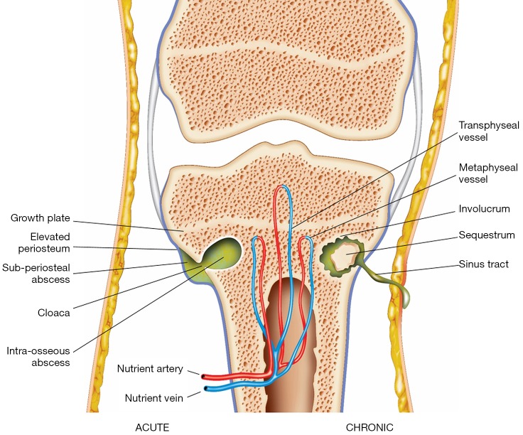
Osteomyelitis is an important cause of morbidity and mortality in children and adults. Imaging plays a crucial role in establishing a timely diagnosis and guiding early management, with the aim of reducing long-term complications. Recognition of the imaging features of osteomyelitis requires a good understanding of its pathogenesis. In this review, the key imaging findings in osteomyelitis are correlated with the underlying pathological processes. There is a particular emphasis on magnetic resonance imaging (MRI), which is the best available imaging modality owing to its high sensitivity for detecting early osteomyelitis, excellent anatomical detail and superior soft tissue resolution. However, other modalities such as nuclear medicine and computed tomography (CT) are also useful in many clinical contexts, and will also be described in this review.

Keywords: Musculoskeletal; infection; magnetic resonance imaging (MRI); radiology.


Conflict of interest statement

Conflicts of Interest: The authors have no conflicts of interest to declare.

Figures

Figure 1
Figure 1
The pathogenesis of osteomyelitis. Metaphyseal vessels contain slow-flowing blood, predisposing to bacterial proliferation. Hence, the metaphysis is a common site for haematogenous osteomyelitis. The growth plate forms a barrier between the metaphyseal and epiphyseal vessels in children over 18 months of age. However, in infants under 18 months and in adults, transphyseal vessels are present which provide a route for infection to communicate between the metaphysis and epiphysis. In acute osteomyelitis, a collection of pus becomes surrounded by granulation tissue and reactive bone, forming an intraosseous abscess. Raised intramedullary pressure secondary to accumulation of pus leads to rupture of the cortex, creating a defect known as a cloaca, which drains pus from the bone to the surrounding tissues. This can cause a subperiosteal abscess with elevation of the periosteum, as well as soft tissue abscesses. In chronic osteomyelitis, disruption of the intraosseous and periosteal blood supply leads to formation of a necrotic bone fragment, known as a sequestrum, which is surrounded by pus and granulation tissue. A reactive shell of new bone forms around the sequestrum and is known as an involucrum. A sinus tract, which drains pus from bone to the skin surface, may be present in both acute and chronic osteomyelitis.
Figure 2
Figure 2
Osteomyelitis in the right foot of a 63-year-old male. (A) The dorso-plantar radiograph shows a periosteal reaction around the 1st metatarsal diaphysis (white arrowheads); (B) short axis coronal short-tau inversion recovery (STIR) image of the same patient demonstrating marked soft tissue oedema surrounding the 1st metatarsal. The periosteum (white arrowheads) is separated from the cortex (white arrow) by high signal material representing pus. There is a defect in the cortex (black arrow), known as a cloaca, that allows pus to drain from the medullary cavity into the subperiosteal space. Compared to the other metatarsals, the medulla (M) of the 1st metatarsal has high signal, consistent with bone marrow oedema.
Figure 3
Figure 3
A 6-year-old girl with no history of trauma presents with pain and swelling in her right knee. (A) The anteroposterior radiograph shows a well-circumscribed lucent lesion with sclerotic margins in the right distal femur metaphysis, suspicious for an intraosseous abscess; (B) coronal STIR image of the right femur shows that the lesion is within the medullary cavity and has high signal (black arrow). The bone marrow of the distal diaphysis and metaphysis has diffuse high signal (white arrow) compared to the mid-diaphysis, representing bone marrow oedema; (C) coronal T1W image shows that the intraosseous lesion has central heterogeneous low signal (black arrow). The bone marrow of the distal diaphysis and metaphysis has diffuse low signal consistent with oedema (white arrow). Note the distinct margin between normal and abnormal marrow, suggestive of bone marrow oedema due to osteomyelitis rather than reactive osteitis; (D) coronal fat-suppressed T1W image after administration of intravenous contrast shows that the lesion has central low signal (black arrow) and peripheral enhancement (white arrowheads). The central low signal represents pus and the peripherally enhancing areas represent hypervascular granulation tissue. This confirms an intraosseous abscess. Its location in the metaphysis is characteristic for haematogenous osteomyelitis.
Figure 4
Figure 4
Chronic osteomyelitis in a 9-year-old boy with a non-united left distal humerus fracture. (A) The lateral radiograph shows marked periosteal thickening (black arrowheads) and a central sclerotic lesion with a lucent rim (black arrow); (B) coronal CT with bone windows shows a sclerotic fragment of bone which is separate from the rest of the humerus (black arrow), consistent with a sequestrum. Cortical thickening is also noted (black arrowheads); this represents an involucrum which is a result of periosteal new bone formation. These findings were not present on initial images taken at the time of the fracture; (C) coronal STIR image shows the low signal sequestrum (black arrow) surrounded by high signal pus and granulation tissue (white arrowheads). There is a sinus tract draining pus to the skin surface (white arrow); (D) axial fat-suppressed T2 image demonstrates that the pus surrounding the sequestrum (black arrow) communicates with the sinus tract (white arrow) via a cloaca (white arrowhead). There is also a soft tissue fluid collection anteromedial to the humerus (black arrowheads). (Images courtesy of Dr. Asif Saifuddin, Royal National Orthopaedic Hospital, Stanmore).
Figure 5
Figure 5
Osteomyelitis in the right tibia of a 44-year-old male. (A) Axial STIR image of both legs. There is high signal in the medullary cavity of the right tibia (M) compared to the normal low signal medulla of the left tibia. This could represent either bone marrow oedema or an intramedullary abscess. There is also circumferential periosteal elevation and high signal (black arrowheads), suggesting a periosteal reaction with a possible subperiosteal abscess. Within the tibial cortex (C), there is a focal area of high signal intensity (white arrow), suspicious for an intraosseous abscess; (B) axial fat-suppressed T1W image following intravenous contrast. The cortical lesion (black arrow) has central low signal and peripheral enhancement, confirming the suspicion of a cortical abscess. There is uniform enhancement of the bone marrow (M) and periosteum (white arrowheads), consistent with bone marrow oedema and a periosteal reaction. The absence of central low signal in these regions excludes intramedullary and subperiosteal abscesses.
Figure 6
Figure 6
A 16-year-old female who stepped on a wooden splinter 2 months earlier, now presents with right foot pain and purulent discharge from the puncture wound. (A) The dorso-plantar radiograph shows sclerosis of the 2nd and 3rd metatarsals, with surrounding periosteal reaction (black arrowheads). The wooden splinter is radiolucent; (B) coronal STIR image shows high signal in the medulla of the 3rd metatarsal (white arrow), consistent with bone marrow oedema; (C) axial STIR image shows that the splinter (black arrow) has become embedded in the 2nd intermetatarsal space. It is surrounded by a high signal fluid collection (white arrowheads) which communicates via a sinus tract (black arrowhead) to the skin surface; (D) axial STIR image at a more distal level showing bone marrow oedema (white arrow), periosteal elevation (white arrowheads) and a sinus tract extending from the bone to the skin surface (black arrow).
Figure 7
Figure 7
Suspected periprosthetic infection in a 72-year-old male with history of bilateral total knee replacements. A combined white cell and marrow scan was performed. (A) Indium 111-labelled white cell scan of the knees demonstrates accumulation of radiolabelled white cells in the left knee between the 3-hour and 24-hour images (black arrows); (B) the technetium 99m-labelled bone marrow scan provides a baseline map of physiological white cell uptake. No tracer uptake is seen in the left knee (white arrows). The discordance between the white cell and marrow scan demonstrates that the white cell uptake is likely to be due to a focus of infection; (C,D) hybrid SPECT/CT imaging fuses functional and anatomical information to enable more accurate localisation of the focus of infection. The discordance between the white cell (white arrows) and marrow scans is again demonstrated, suggestive of periprosthetic infection.
Figure 8
Figure 8
Osteomyelitis may be associated with soft tissue collections which can be seen on ultrasound. (A) Transverse section ultrasound image demonstrating a well-defined complex fluid collection which has an irregular thick wall (white arrowheads) and a hyperechoic septation (white arrow); (B) percutaneous needle aspiration of the fluid collection was performed (black arrowheads). Culture of the aspirate grew Staphylococcus aureus.
Figure 9
Figure 9
Flow chart for imaging modality choice in osteomyelitis. A plain radiograph should always be obtained first to exclude fractures. Unless contraindicated, an MRI should then be performed as it is the best currently available modality for establishing the diagnosis of osteomyelitis. If MRI is contraindicated, CT or nuclear medicine studies can be obtained, although these tests are of limited sensitivity and specificity compared to MRI.

References

    1. Lew DP, Waldvogel FA. Osteomyelitis. Lancet 2004;364:369-79. 10.1016/S0140-6736(04)16727-5 - DOI - PubMed
    1. Jaramillo D. Infection: musculoskeletal. Pediatr Radiol 2011;41 Suppl 1:S127-34. 10.1007/s00247-011-2001-y - DOI - PubMed
    1. Arnold SR, Elias D, Buckingham SC, Thomas ED, Novais E, Arkader A, Howard C. Changing patterns of acute hematogenous osteomyelitis and septic arthritis: emergence of community-associated methicillin-resistant Staphylococcus aureus. J Pediatr Orthop 2006;26:703-8. 10.1097/01.bpo.0000242431.91489.b4 - DOI - PubMed
    1. Gafur OA, Copley LA, Hollmig ST, Browne RH, Thornton LA, Crawford SE. The impact of the current epidemiology of pediatric musculoskeletal infection on evaluation and treatment guidelines. J Pediatr Orthop 2008;28:777-85. 10.1097/BPO.0b013e318186eb4b - DOI - PubMed
    1. Pugmire BS, Shailam R, Gee MS. Role of MRI in the diagnosis and treatment of osteomyelitis in pediatric patients. World J Radiol 2014;6:530-7. 10.4329/wjr.v6.i8.530 - DOI - PMC - PubMed

LinkOut - more resources