

Synchronous Kimura lesions at two different sites-a diagnostic dilemma!
- PMID: 27190775
- PMCID: PMC4858467
- DOI: 10.21037/qims.2015.08.04
Synchronous Kimura lesions at two different sites-a diagnostic dilemma!
Abstract
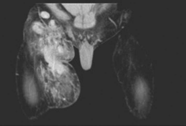
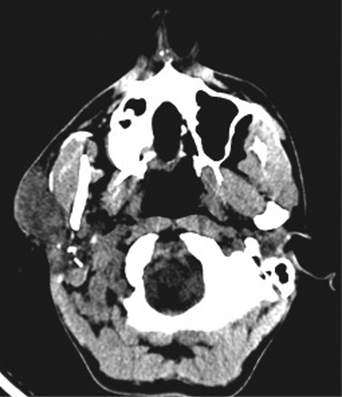
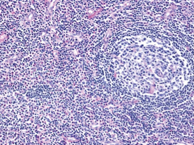
Kimura disease (KD) is a rare, chronic, benign inflammatory disorder of unknown etiology mimicking neoplastic disease and is characterized by multiple subcutaneous nodules and masses, primarily in the cervical region, accompanied by peripheral eosinophilia and lymphadenopathy. A 35-year-old male presented with a 2-year history of swelling in the right preauricular region and right thigh. Investigations showed a peripheral eosinophilia. CT of cervical region revealed parotid neoplasm and a fine needle aspiration was inconclusive. The thigh swelling felt vascular and a CT angiogram was done which revealed a diffuse vascular lesion. A superficial parotidectomy and an excision of the right thigh swelling were done. Histopathologies of both specimens were reported to have features suggestive of KD. The patient was evaluated for systemic manifestations and found to have no abnormalities. The patient has been disease free on follow-up after 2 years. This case is being presented for the rarity of its incidence and the nature of its presentation.
Keywords: Kimura; eosinophilia; lymphadenopathy; parotidectomy.
Conflict of interest statement
Figures
Similar articles
-
Kimura's Disease without Peripheral Eosinophilia: An Unusual and Challenging Case Simulating Venous Malformation on Imaging Studies-Case Report and Review of literature.J Clin Diagn Res. 2017 Jun;11(6):ME01-ME04. doi: 10.7860/JCDR/2017/28603.10063. Epub 2017 Jun 1. J Clin Diagn Res. 2017. PMID: 28764210 Free PMC article. Review.
-
Kimura disease: A rare case affecting the inguinal lymph node.Int J Surg Case Rep. 2025 Jun;131:111332. doi: 10.1016/j.ijscr.2025.111332. Epub 2025 Apr 21. Int J Surg Case Rep. 2025. PMID: 40334445 Free PMC article.
-
Kimura Disease: An Unusual Presentation of Parotid Mass in a Sickle Cell Disease Patient.Cureus. 2022 May 6;14(5):e24787. doi: 10.7759/cureus.24787. eCollection 2022 May. Cureus. 2022. PMID: 35676993 Free PMC article.
-
Superficial Parotidectomy Plane for Debulking Surgery in Kimura Disease.J Craniofac Surg. 2017 May;28(3):e207-e208. doi: 10.1097/SCS.0000000000003387. J Craniofac Surg. 2017. PMID: 28468187
-
Kimura disease: a clinicopathologic study of 21 cases.Am J Surg Pathol. 2004 Apr;28(4):505-13. doi: 10.1097/00000478-200404000-00010. Am J Surg Pathol. 2004. PMID: 15087670 Review.
Cited by
-
Kimura's Disease without Peripheral Eosinophilia: An Unusual and Challenging Case Simulating Venous Malformation on Imaging Studies-Case Report and Review of literature.J Clin Diagn Res. 2017 Jun;11(6):ME01-ME04. doi: 10.7860/JCDR/2017/28603.10063. Epub 2017 Jun 1. J Clin Diagn Res. 2017. PMID: 28764210 Free PMC article. Review.
References
-
- Kawada A. Kimura’s disease. Demonstration of the disease and its differential diagnosis. Hautarzt 1976;27:309-17. - PubMed
Publication types
LinkOut - more resources
Full Text Sources
Other Literature Sources