

The Serum Levels of the Soluble Factors sCD40L and CXCL1 Are Not Indicative of Endometriosis
- PMID: 27190986
- PMCID: PMC4852116
- DOI: 10.1155/2016/2857161
The Serum Levels of the Soluble Factors sCD40L and CXCL1 Are Not Indicative of Endometriosis
Abstract
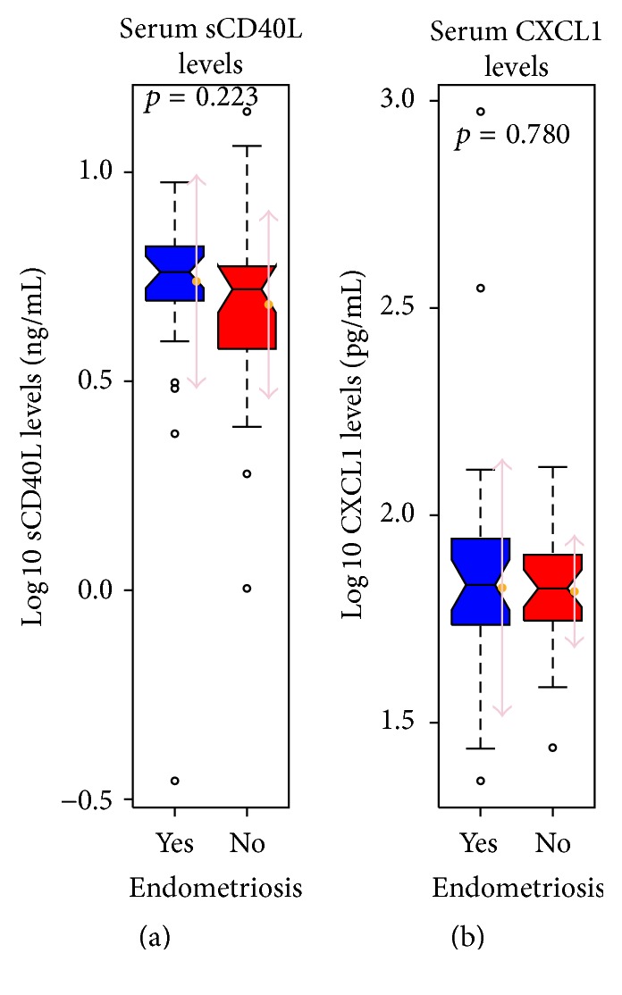
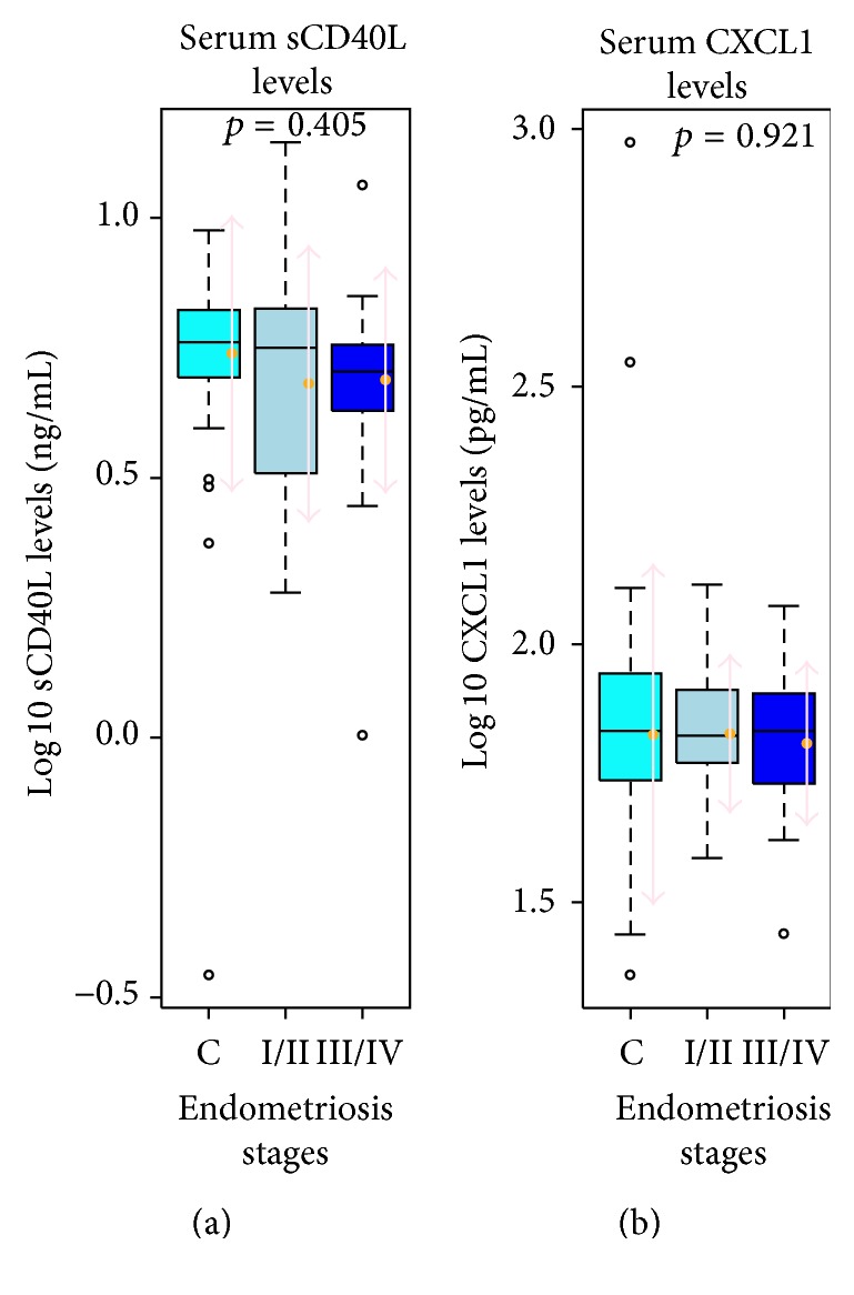
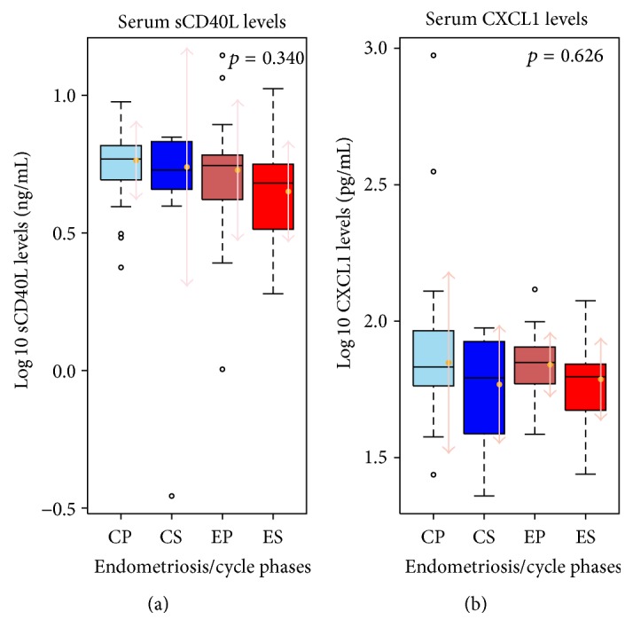
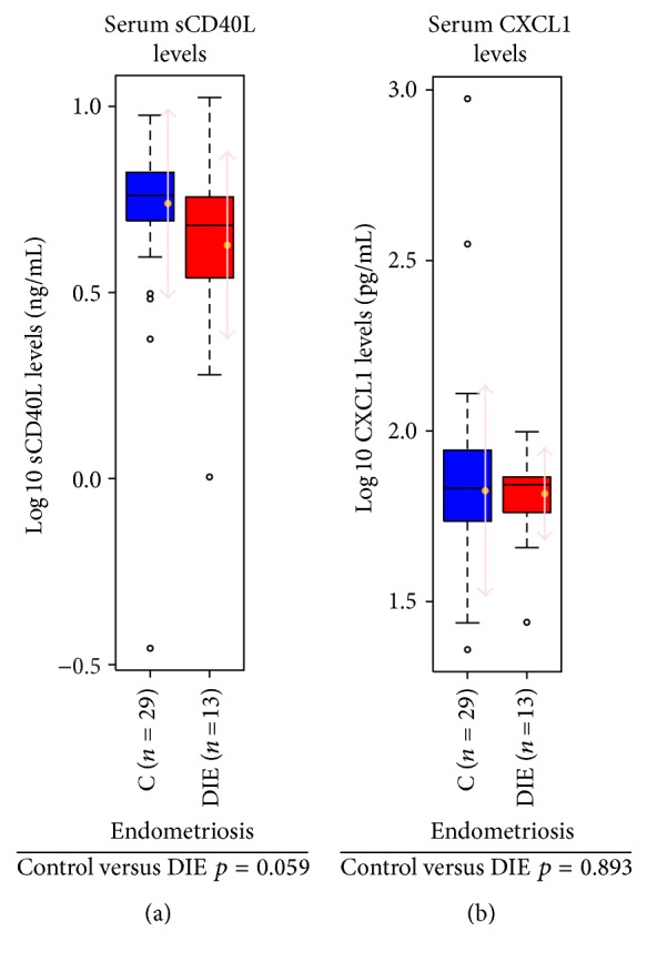
Endometriosis is a benign but troublesome gynecological condition, characterized by endometrial-like tissue outside the uterine cavity. Lately, the discovery and validation of noninvasive diagnostic biomarkers for endometriosis is one of the main priorities in the field. As the disease elicits a chronic inflammatory reaction, we focused our interest on two factors well known to be involved in inflammation and neoplastic processes, namely, soluble CD40 Ligand and CXCL1, and asked whether differences in the serum levels of sCD40L and CXCL1 in endometriosis patients versus controls can serve as noninvasive disease markers. A total of n = 60 women were included in the study, 31 endometriosis patients and 29 controls, and the serum levels of sCD40L and CXCL1 were measured by enzyme-linked immunosorbent assay. Overall, there were no statistically significant differences in the levels of expression of both sCD40L and CXCL1 between patients and controls. This study adds useful clinical data showing that the serum levels of the soluble factors sCD40L and CXCL1 are not associated with endometriosis and are not suitable as biomarkers for disease diagnosis. However, we found a trend toward lower levels of sCD40L in the deep infiltrating endometriosis subgroup making it a potentially interesting target worth further investigation.
Figures
Similar articles
-
The levels of circulating markers of atherosclerosis and inflammation in subjects with different degrees of body mass index: Soluble CD40 ligand and high-sensitivity C-reactive protein.Thromb Res. 2007;119(1):79-84. doi: 10.1016/j.thromres.2005.12.019. Epub 2006 Feb 14. Thromb Res. 2007. PMID: 16476470
-
Elevated circulating soluble CD40 ligand in patients with mixed connective tissue disease.Clin Rheumatol. 2003 Feb;22(1):37-9. doi: 10.1007/s10067-002-0669-y. Clin Rheumatol. 2003. PMID: 12605316
-
Increased plasma soluble CD40 ligand concentration in pelvic inflammatory disease.Clin Chim Acta. 2015 Jan 1;438:236-40. doi: 10.1016/j.cca.2014.08.030. Epub 2014 Sep 2. Clin Chim Acta. 2015. PMID: 25192781
-
Plasma levels of soluble CD40 ligand are elevated in inflammatory bowel diseases.Int J Colorectal Dis. 2003 Mar;18(2):142-7. doi: 10.1007/s00384-002-0425-4. Epub 2002 Sep 5. Int J Colorectal Dis. 2003. PMID: 12548417
-
Plasma matrix metalloproteinase-9, tissue inhibitor of metalloproteinase-2, and CD40 ligand levels in patients with stable coronary artery disease.Am J Cardiol. 2005 Aug 1;96(3):339-45. doi: 10.1016/j.amjcard.2005.03.072. Am J Cardiol. 2005. PMID: 16054454
Cited by
-
Chemokine expression in patients with ovarian cancer or benign ovarian tumors.Arch Med Sci. 2021 Mar 21;18(3):682-689. doi: 10.5114/aoms/110672. eCollection 2022. Arch Med Sci. 2021. PMID: 35591828 Free PMC article.
-
Challenges in uncovering non-invasive biomarkers of endometriosis.Exp Biol Med (Maywood). 2020 Mar;245(5):437-447. doi: 10.1177/1535370220903270. Epub 2020 Feb 4. Exp Biol Med (Maywood). 2020. PMID: 32019326 Free PMC article. Review.
-
The Importance of CXCL1 in the Physiological State and in Noncancer Diseases of the Oral Cavity and Abdominal Organs.Int J Mol Sci. 2022 Jun 28;23(13):7151. doi: 10.3390/ijms23137151. Int J Mol Sci. 2022. PMID: 35806156 Free PMC article. Review.
References
MeSH terms
Substances
LinkOut - more resources
Full Text Sources
Other Literature Sources
Medical
Research Materials
