

Arylfluorosulfates Inactivate Intracellular Lipid Binding Protein(s) through Chemoselective SuFEx Reaction with a Binding Site Tyr Residue
- PMID: 27191344
- PMCID: PMC4909538
- DOI: 10.1021/jacs.6b02960
Arylfluorosulfates Inactivate Intracellular Lipid Binding Protein(s) through Chemoselective SuFEx Reaction with a Binding Site Tyr Residue
Abstract
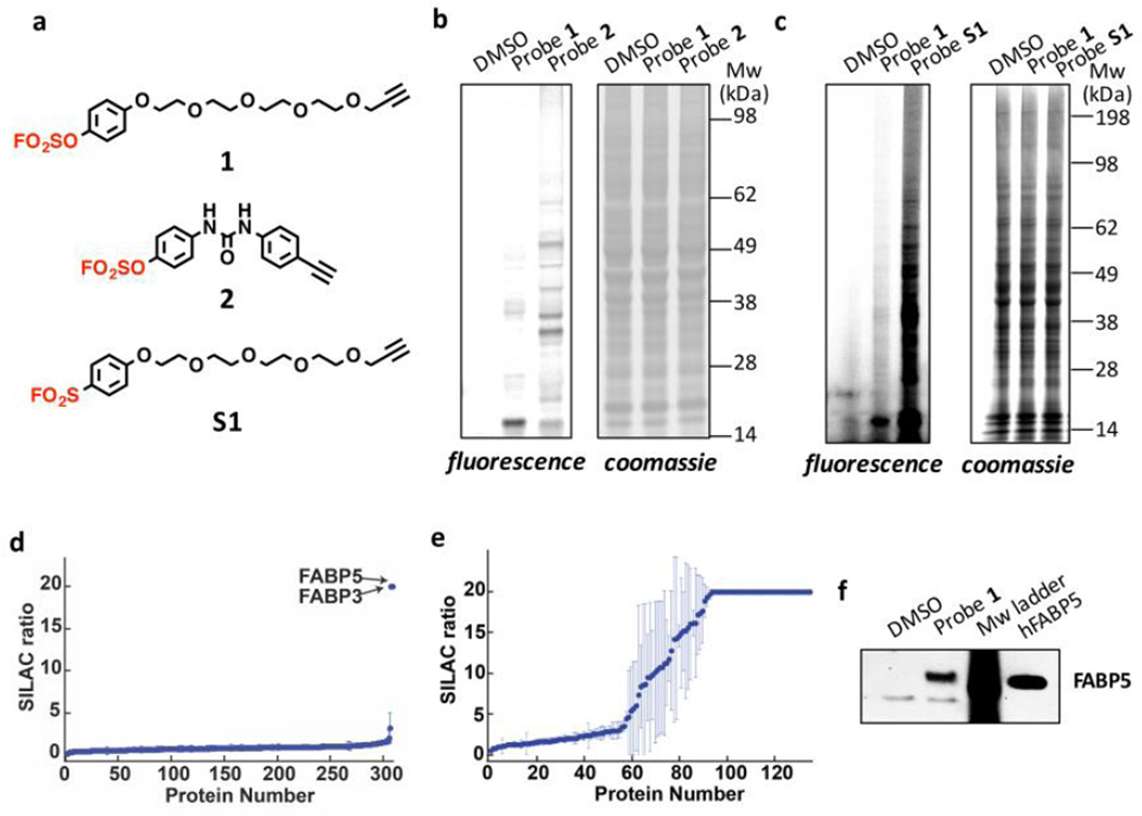
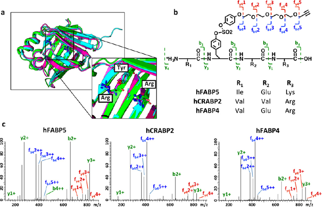
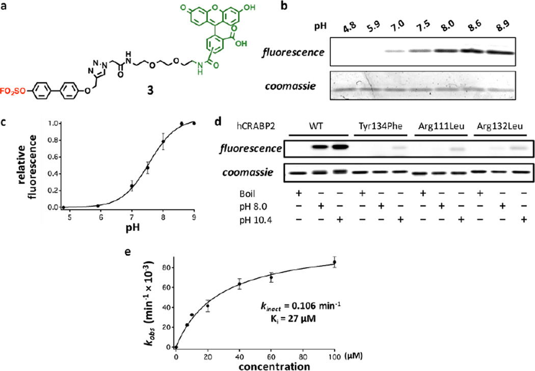
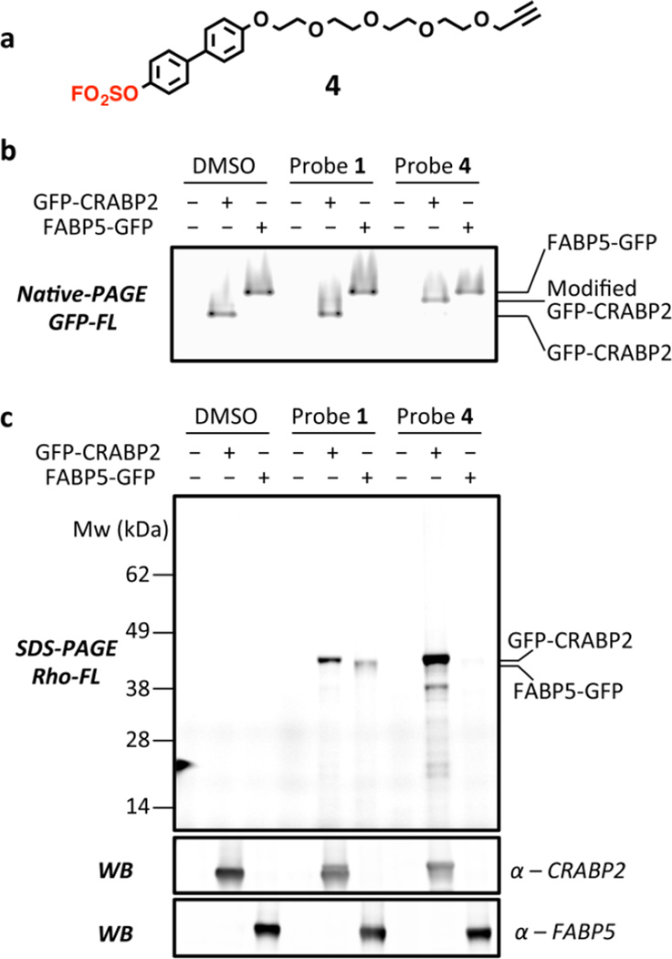
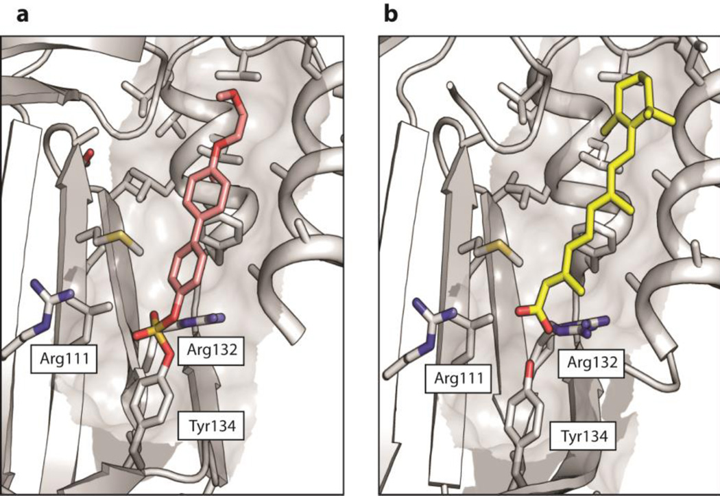
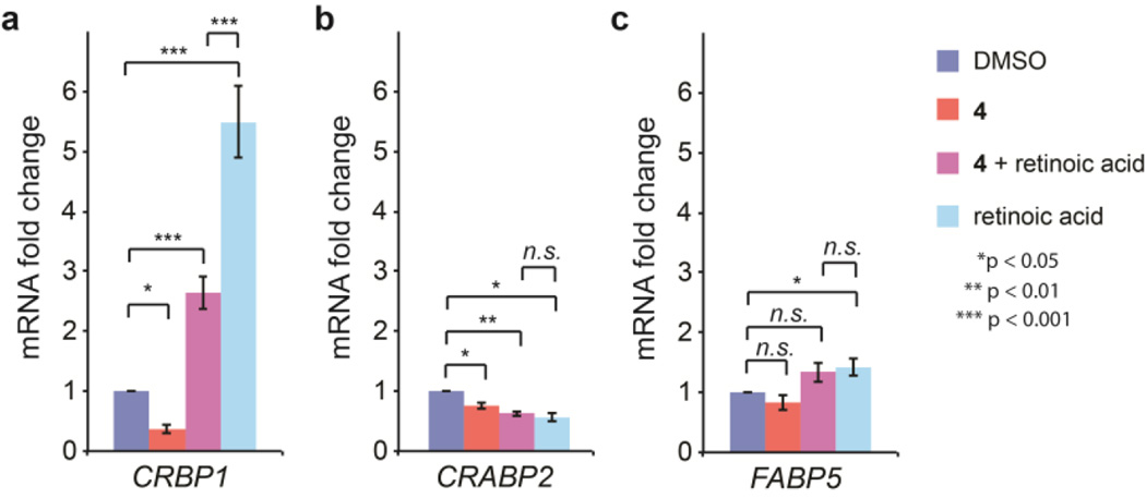
Arylfluorosulfates have appeared only rarely in the literature and have not been explored as probes for covalent conjugation to proteins, possibly because they were assumed to possess high reactivity, as with other sulfur(VI) halides. However, we find that arylfluorosulfates become reactive only under certain circumstances, e.g., when fluoride displacement by a nucleophile is facilitated. Herein, we explore the reactivity of structurally simple arylfluorosulfates toward the proteome of human cells. We demonstrate that the protein reactivity of arylfluorosulfates is lower than that of the corresponding aryl sulfonyl fluorides, which are better characterized with regard to proteome reactivity. We discovered that simple hydrophobic arylfluorosulfates selectively react with a few members of the intracellular lipid binding protein (iLBP) family. A central function of iLBPs is to deliver small-molecule ligands to nuclear hormone receptors. Arylfluorosulfate probe 1 reacts with a conserved tyrosine residue in the ligand-binding site of a subset of iLBPs. Arylfluorosulfate probes 3 and 4, featuring a biphenyl core, very selectively and efficiently modify cellular retinoic acid binding protein 2 (CRABP2), both in vitro and in living cells. The X-ray crystal structure of the CRABP2-4 conjugate, when considered together with binding site mutagenesis experiments, provides insight into how CRABP2 might activate arylfluorosulfates toward site-specific reaction. Treatment of breast cancer cells with probe 4 attenuates nuclear hormone receptor activity mediated by retinoic acid, an endogenous client lipid of CRABP2. Our findings demonstrate that arylfluorosulfates can selectively target single iLBPs, making them useful for understanding iLBP function.
Figures
Similar articles
-
Novel Approaches to Access Arylfluorosulfates and Sulfamoyl Fluorides Based on Sulfur (VI) Fluoride Exchange.Curr Protoc Chem Biol. 2019 Jun;11(2):e64. doi: 10.1002/cpch.64. Epub 2019 Feb 28. Curr Protoc Chem Biol. 2019. PMID: 30816629 Free PMC article.
-
The Role of Aromatic-Aromatic Interactions in Strand-Strand Stabilization of β-Sheets.J Mol Biol. 2013 Sep 23;425(18):3522-35. doi: 10.1016/j.jmb.2013.06.030. Epub 2013 Jun 28. J Mol Biol. 2013. PMID: 23810905 Free PMC article.
-
Arylfluorosulfate-Based Electrophiles for Covalent Protein Labeling: A New Addition to the Arsenal.Angew Chem Int Ed Engl. 2019 Jan 21;58(4):957-966. doi: 10.1002/anie.201806037. Epub 2018 Oct 26. Angew Chem Int Ed Engl. 2019. PMID: 30024079 Free PMC article. Review.
-
Sequence and structural analysis of cellular retinoic acid-binding proteins reveals a network of conserved hydrophobic interactions.Proteins. 2004 Feb 1;54(2):179-94. doi: 10.1002/prot.10520. Proteins. 2004. PMID: 14696180
-
The role of dynamics in modulating ligand exchange in intracellular lipid binding proteins.Biochim Biophys Acta. 2014 Jul;1844(7):1268-78. doi: 10.1016/j.bbapap.2014.04.011. Epub 2014 Apr 21. Biochim Biophys Acta. 2014. PMID: 24768771 Review.
Cited by
-
Lessons in Organic Fluorescent Probe Discovery.Chembiochem. 2021 Nov 16;22(22):3109-3139. doi: 10.1002/cbic.202100171. Epub 2021 Jun 23. Chembiochem. 2021. PMID: 34062039 Free PMC article. Review.
-
[Advances in applications of activity-based chemical probes in the characterization of amino acid reactivities].Se Pu. 2023 Jan;41(1):14-23. doi: 10.3724/SP.J.1123.2022.05013. Se Pu. 2023. PMID: 36633073 Free PMC article. Review. Chinese.
-
A practical fluorosulfonylating platform via photocatalytic imidazolium-based SO2F radical reagent.Nat Commun. 2022 Jun 18;13(1):3515. doi: 10.1038/s41467-022-31296-2. Nat Commun. 2022. PMID: 35717500 Free PMC article.
-
Liganding Functional Tyrosine Sites on Proteins Using Sulfur-Triazole Exchange Chemistry.J Am Chem Soc. 2020 May 6;142(18):8270-8280. doi: 10.1021/jacs.0c00648. Epub 2020 Apr 24. J Am Chem Soc. 2020. PMID: 32329615 Free PMC article.
-
Tyrosine bioconjugation - an emergent alternative.Org Biomol Chem. 2020 Nov 28;18(44):9018-9028. doi: 10.1039/d0ob01912g. Epub 2020 Nov 3. Org Biomol Chem. 2020. PMID: 33141139 Free PMC article. Review.
References
-
- Dong J, Krasnova L, Finn MG, Sharpless KB. Angew Chem Int Ed Engl. 2014;53:9430. - PubMed
-
- Baker BR. Annu Rev Pharmacol. 1970;10:35. - PubMed
-
- Baker BR. Ann N Y Acad Sci. 1971;186:214. - PubMed
-
- Baker BR, Erickson EH. J Med Chem. 1968;11:245. - PubMed
-
- Baker BR, Hurlbut JA. J Med Chem. 1968;11:233. - PubMed
Publication types
MeSH terms
Substances
Grants and funding
LinkOut - more resources
Full Text Sources
Other Literature Sources
Research Materials
Miscellaneous
