. 2016 May 18;11(5):e0155831.
doi: 10.1371/journal.pone.0155831. eCollection 2016.

PEGylation of Truncated Streptokinase Leads to Formulation of a Useful Drug with Ameliorated Attributes

Affiliations

PEGylation of Truncated Streptokinase Leads to Formulation of a Useful Drug with Ameliorated Attributes

Pooja Sawhney et al. PLoS One. .

Abstract

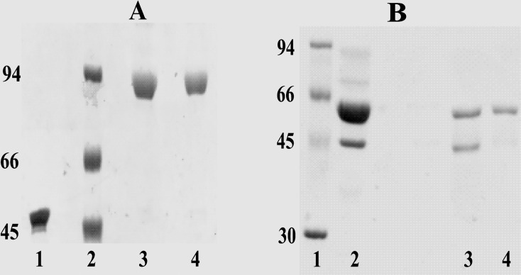
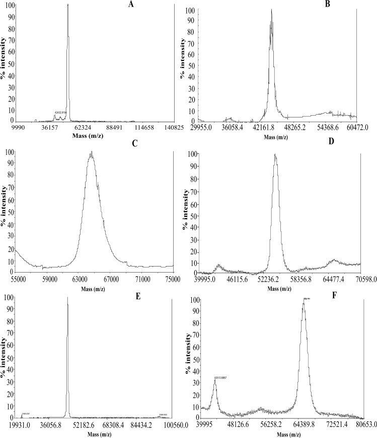
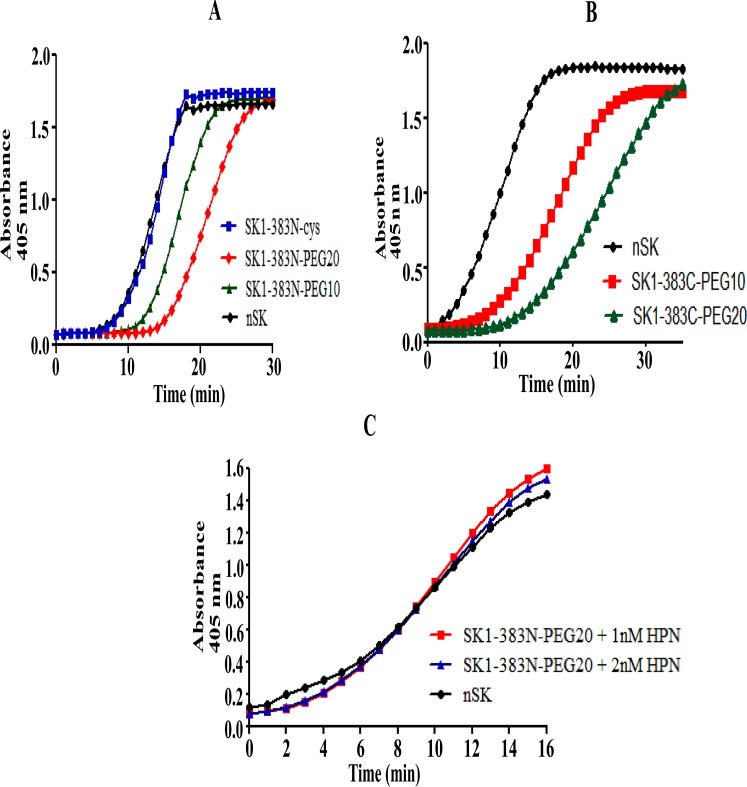
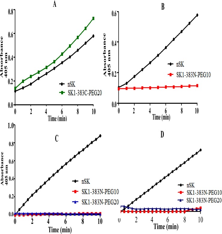
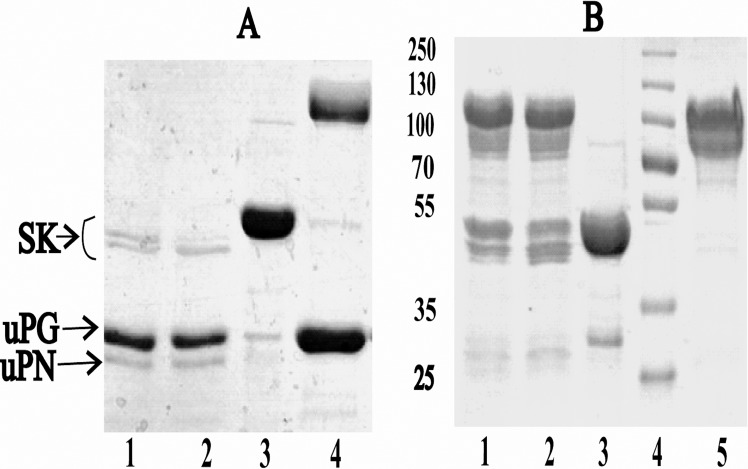
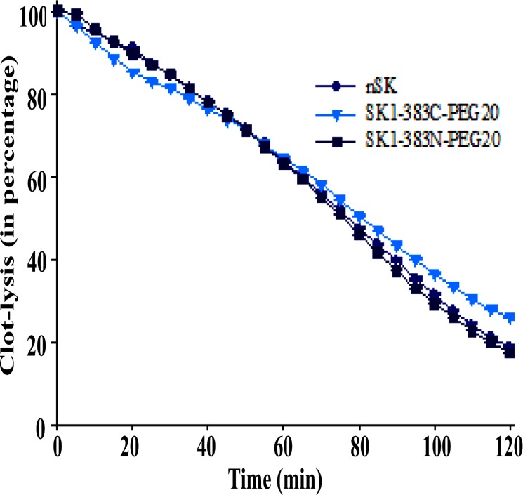
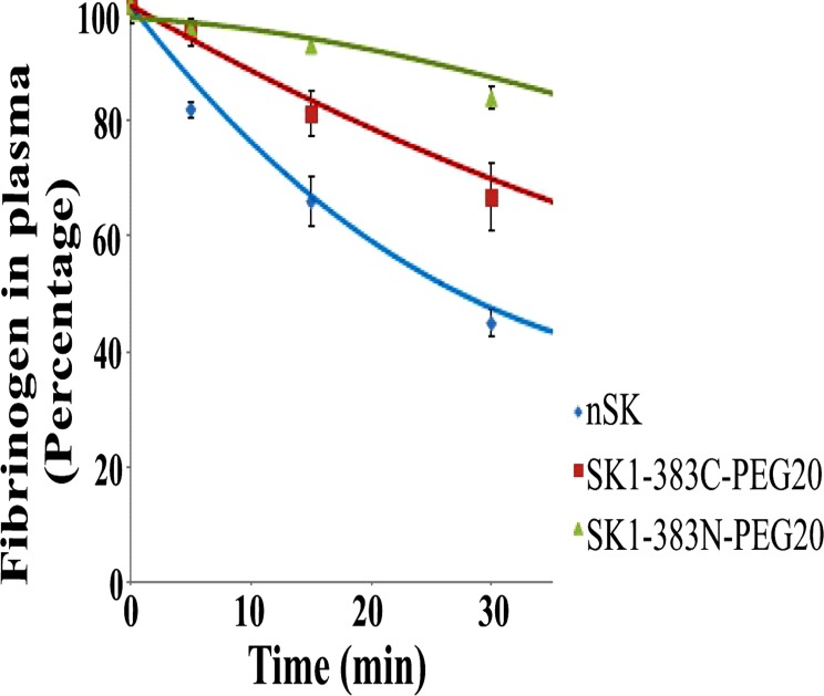
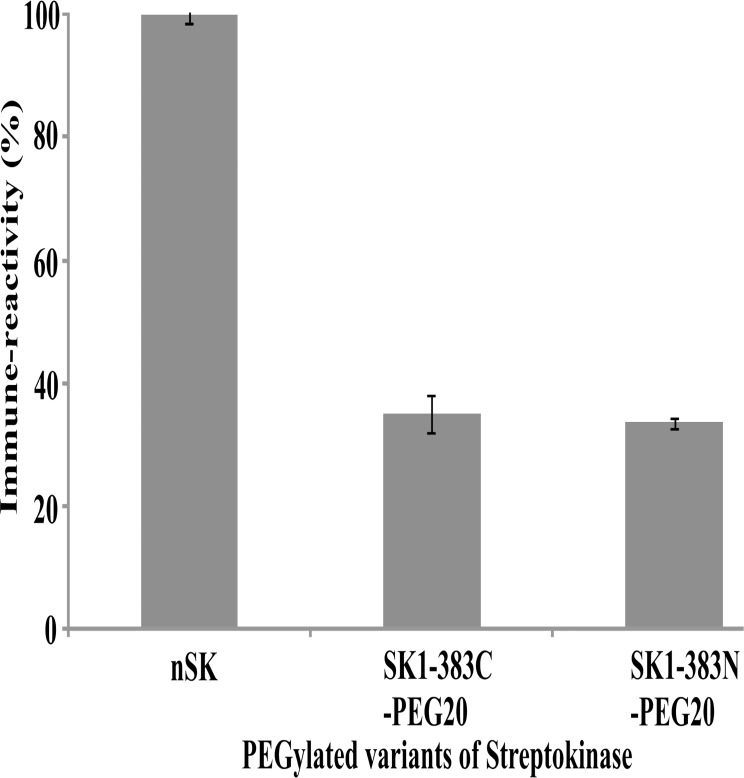
Streptokinase (SK) remains a favored thrombolytic agent in the developing world as compared to the nearly 10-fold more expensive human tissue-plasminogen activator (tPA) for the dissolution of pathological fibrin clots in myocardial infarction. However, unlike the latter, SK induces systemic activation of plasmin which results in a greater risk of hemorrhage. Being of bacterial origin, it elicits generation of unwanted antibody and has a relatively short half-life in vivo that needs to be addressed to make it more efficacious clinically. In order to address these lacunae, in the present study we have incorporated cysteine residues specifically at the N- and C-termini of partially truncated SK and these were then PEGylated successfully. Some of the obtained derivatives displayed enhanced plasmin resistance, longer half-life (upto several hours), improved fibrin clot-specificity and reduced immune-reactivity as compared to the native SK (nSK). This paves the way for devising next-generation SK-based thrombolytic agent/s that besides being fibrin clot-specific are endowed with an improved efficacy by virtue of an extended in vivo half-life.


Conflict of interest statement

Competing Interests: The authors have declared that no competing interests exist.

Figures

Fig 1
Fig 1. SDS-PAGE analysis of the SK-PEGylated protein.
(A) SDS-PAGE analysis of purified SK1-383C-PEG20 (C-terminal truncated cysteine mutant PEGylated with 20KDa moiety) derivative. Lane 1: unpegylated protein; lane 2: molecular weight markers; lanes 3 and 4: Fractions 1 and 2 of purified SK1-383C-PEG20, through gel filtration chromatography. (B) Electrophoresis of SK1-383N-PEG10 (N-terminal cysteine mutant PEGylated with 10KDa moiety) SK derivative stained with coomassie. Lane 1: molecular weight markers; lane 2: PEGylation reaction mixture; lane 3: PEGylated protein after anion-exchange chromatography; lane 4: PEGylated protein after gel filtration chromatography.
Fig 2
Fig 2. Determination of molecular weight of PEGylated derivatives by mass-spectrometry (MALDI-TOF).
Molecular weight determined by MALDI-TOF of the PEGylated and unPEGylated-SK mutants were in agreement with their calculated molecular weight. (A) SK1-383N-PEG10 (B) SK1-383C-cys (C) SK1-383N-PEG20 (D) SK1-383C-PEG10 (E) nSK (F) SK1-383C-PEG20.
Fig 3
Fig 3. Depiction of time-course activation of HPG by nSK, truncated SK and PEGylated SK and PN-dependency of PEG-SK derivatives for the activation of HPG.
(A) nSK/truncated SK/PEGylated-SK (0.5 nM conc. each) were added separately in microtiter plate wells, containing HPG and chromozyme® PL. The progress curves of activation of HPG were measured spectrophotometrically at 405nm over time. The graph shows nSK (black color circles)SK1-383N-cys (blue rectangles), SK1-383N-PEG10 (green triangles), SK1-383N-PEG20 (red diamonds). (B) Graph represents activation of HPG by nSK/SK1-383C-PEG derivatives at 1 nM conc. each in the assay. nSK (black circles), SK1-383C-PEG10 (red rectangles), SK1-383C-PEG20 (green triangles). (C) Plasmin (1 nM or 2 nM) was incubated in a well containing nSK/truncated PEG-SK derivative, HPG and chromozyme ® PL. Graph depicts native SK (black circles), SK1-383N-PEG20 containing 1 nM HPN (red rectangles), SK1-383N-PEG20 containing 2 nM HPN (blue triangles). Statistical analysis showed significant difference (p-value < 0.0001, n = 10, one-way ANOVA test).
Fig 4
Fig 4. Amidolytic activity of nSK/truncated PEG-SK complexes with HPG/μPG at 37°C and 4°C.
Pre-incubated HPG/uPG with nSK/truncated PEG-SK derivatives were added into the cuvette containing chromozyme ® PL. Amidolytic activity generation were monitored by change in absorbance at A405 by hydrolysis of the substrate S-2251. (A) Graph displays amidolytic activation by native SK (black circles) and SK1-383C-PEG20 (green rectangles) in complex with μPG at 37°C. (B) Graph depicts the effect of conjugation of PEG moiety at N-terminal of derivative SK1-383N-PEG10 (red rectangles) by blocking its amidolytic activation in comparison with nSK (black circles), after making complex with μPG, at 37°C. (C) Graph displays the blocking of pathway I by N-terminally PEGylated SK truncated derivatives i.e. SK1-383N-PEG10 (red rectangles), SK1-383N-PEG20 (blue triangles) in comparison with nSK (black circles), when in complex with uPG, at 4°C. (D) Graph depicts no amidolytic activation by SK1-383N-PEG10 (red rectangles) and SK1-383N-PEG20 (blue triangles) in comparison with nSK (black circles), after making 1:1 complex with HPG, at 4°C. Statistical analysis showed significant difference (p-value < 0.0001, n = 6, one-way ANOVA test).
Fig 5
Fig 5. SDS-PAGE analysis of the HPG/μPG-nSK/PEG-SK complexes.
Native SK/PEG-SK was incubated with HPG/μPG to form 1:1 complex at 5 μM concentration. After 3 and 10 min, each of these complexes were subjected to SDS-PAGE under reducing conditions. (A) Gel demonstrates the fragmentation of nSK after complex formation with μPG while no effect was noticed with PEG-SK derivatives. Lane 1: nSK-μPG complex (3 min); lane 2: nSK-μPG complex (10 min); lane 3: Streptokinase alone; lane 4: SK1-383N-PEG20-μPG (10 min). (B) SDS-PAGE indicates the blockage of pathway I when in complex with HPG. Lane 1: nSK-HPG complex (3 min); lane 2: nSK-HPG complex (10 min); lane 3: Streptokinase alone; lane 4: Molecular weight marker; lane 5: SK1-383N-PEG20-HPG (10 min).
Fig 6
Fig 6. Demonstration of clot-lysis profile of PEG-SK variants in comparison with nSK at 5 nM concentration.
A 100 μl of clot was incubated with nSK or its PEGylated derivatives in a microtitre plate at 37°C. Lysis of the clot was measured with time at A405 nm on ELISA plate reader. Graph represents the lysis of clot by SK1-383N-PEG20 (dark blue rectangles) and SK1-383C-PEG20 (blue triangles) in comparison with nSK (black circles). Experiment was performed in triplicates and standard deviation is shown with error bars.
Fig 7
Fig 7. Determination of fibrinogen (in percentage) in the presence of native SK and PEG-SK variants in plasma.
25 nM concentration of nSK/truncated PEGylated derivatives were incubated with plasma and suitable volume of aliquots were removed after every 10 min for determination of the residual fibrinogen content. Graph represents the rate of fibrinogen degradation (in percentage) with time at 25 nM concentration by nSK (blue diamonds), SK1-383N-PEG20 (green triangles) and SK1-383C-PEG20 (red rectangles). Experiment was performed in triplicates and standard deviation is shown with error bars.
Fig 8
Fig 8. Demonstration of the relative reactivity of PEGylated SK truncated variants with rabbit polysera.
The reactivity of nSK and PEGylated SK truncated variants were determined against rabbit polysera and expressed as percentage relative to that displayed by the former (taken as 100 percent reactivity). ELISA plates were coated with nSK and competition against polysera by PEGylated and unPEGylated (indicated on abscissa of each graph) proteins were assessed separately (see Materials and Methods section). Experiment was performed in triplicates and standard deviation is shown with error bars.

References

    1. Tillett WS, Garner RL. The Fibrinolytic Activity of Hemolytic Streptococci. J Exp Med. 1933;58:485–502. - PMC - PubMed
    1. Collen D. Coronary thrombolysis: streptokinase or recombinant tissue-type plasminogen activator? Annals of internal medicine. 1990;112(7):529–38. - PubMed
    1. Sikri N, Bardia A. A history of streptokinase use in acute myocardial infarction. Texas Heart Institute Journal. 2007;34(3):318 - PMC - PubMed
    1. Reddy KNN, Markus G. Mechanism of activation of human plasminogen by streptokinase Presence of active center in streptokinase-plasminogen complex. Journal of Biological Chemistry. 1972;247(6):1683–91. - PubMed
    1. Aneja R, Datt M, Singh B, Kumar S, Sahni G. Identification of a new exosite involved in catalytic turnover by the streptokinase-plasmin activator complex during human plasminogen activation. Journal of Biological Chemistry. 2009;284(47):32642–50. 10.1074/jbc.M109.046573 - DOI - PMC - PubMed