

Wnt Signaling Alteration in the Spinal Cord of Amyotrophic Lateral Sclerosis Transgenic Mice: Special Focus on Frizzled-5 Cellular Expression Pattern
- PMID: 27192435
- PMCID: PMC4871528
- DOI: 10.1371/journal.pone.0155867
Wnt Signaling Alteration in the Spinal Cord of Amyotrophic Lateral Sclerosis Transgenic Mice: Special Focus on Frizzled-5 Cellular Expression Pattern
Abstract
Background: Amyotrophic lateral sclerosis is a chronic neurodegenerative disease characterized by progressive paralysis due to degeneration of motor neurons by unknown causes. Recent evidence shows that Wnt signaling is involved in neurodegenerative processes, including Amyotrophic Lateral Sclerosis. However, to date, little is known regarding the expression of Wnt signaling components in this fatal condition. In the present study we used transgenic SOD1G93A mice to evaluate the expression of several Wnt signaling components, with special focus on Frizzled-5 cellular expression alteration along disease progression.
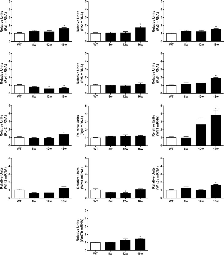
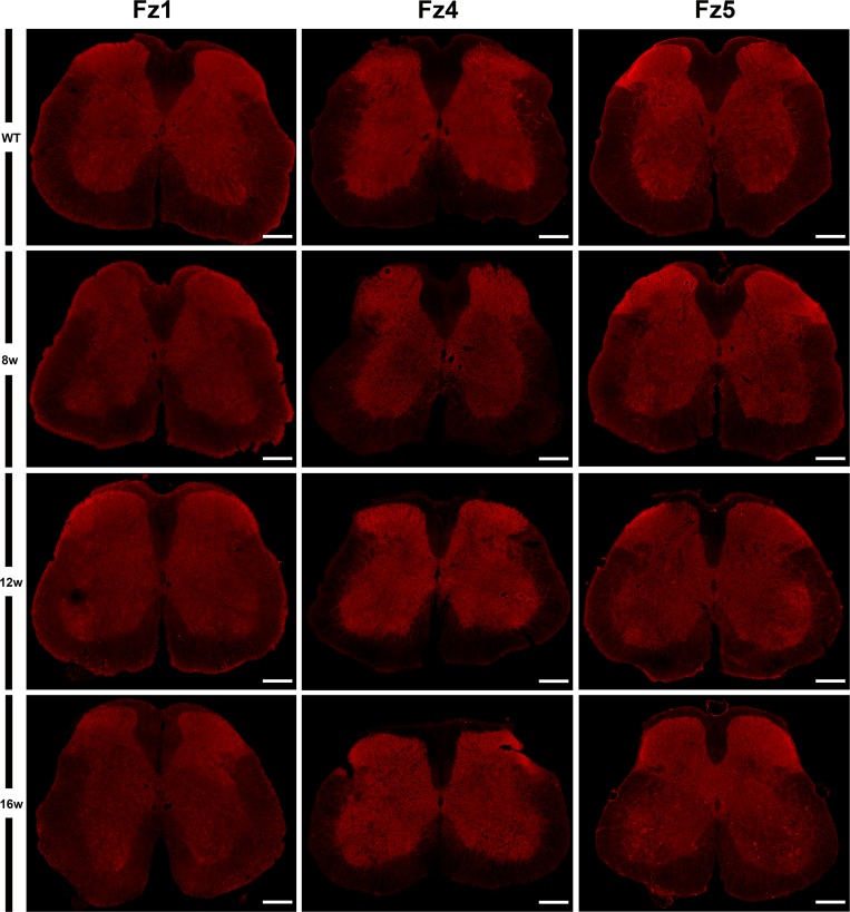
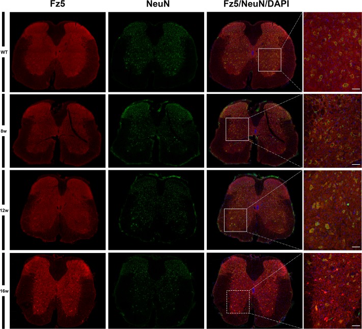
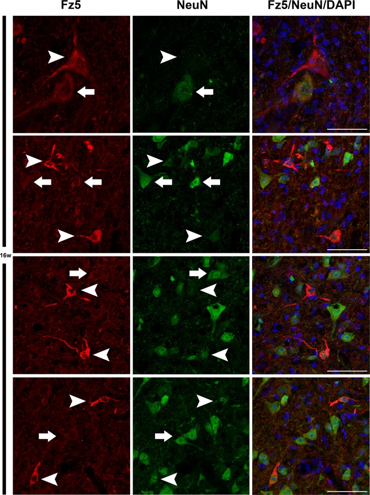
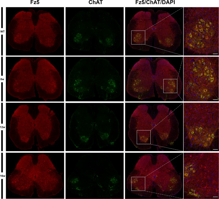
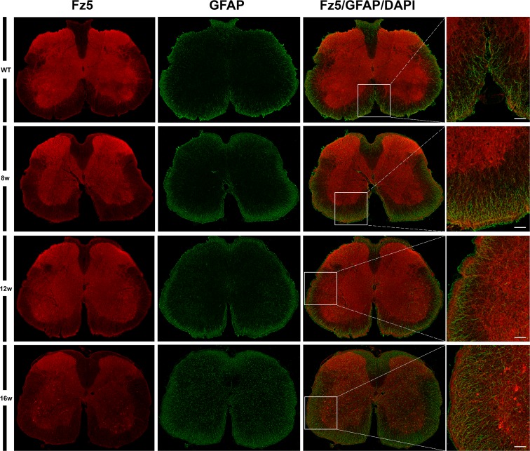
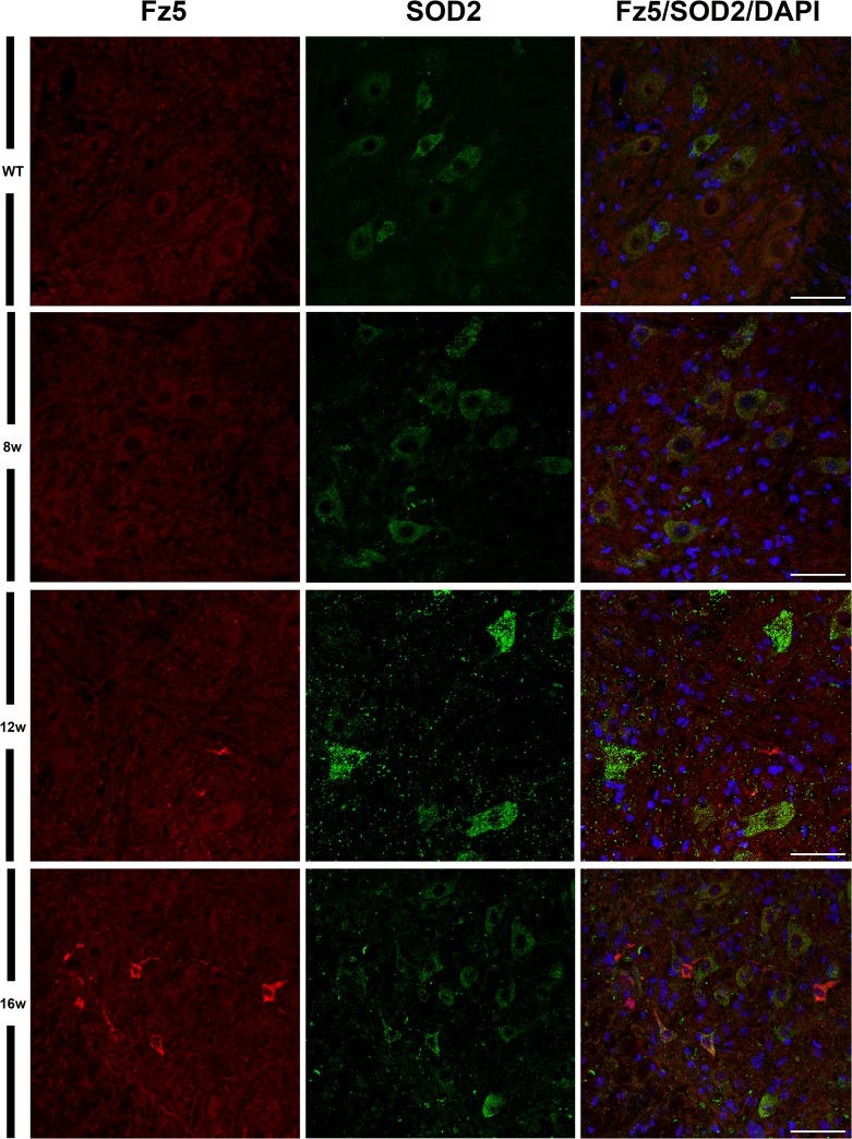
Findings: Based on previous studies demonstrating the expression of Wnts and their transcriptional regulation during Amyotrophic lateral sclerosis development, we have analyzed the mRNA expression of several Wnt signaling components in the spinal cord of SOD1G93A transgenic mice at different stages of the disease by using real time quantitative PCR analysis. Strikingly, one of the molecules that seemed not to be altered at mRNA level, Frizzled-5, showed a clear up-regulation at late stages in neurons, as evidenced by immunofluorescence assays. Moreover, increased Frizzled-5 appears to correlate with a decrease in NeuN signal in these cells, suggesting a correlation between neuronal affectation and the increased expression of this receptor.
Conclusions: Our data suggest the involvement of Wnt signaling pathways in the pathophysiology of Amyotrophic Lateral Sclerosis and, more specifically, the implication of Frizzled-5 receptor in the response of neuronal cells against neurodegeneration. Nevertheless, further experimental studies are needed to shed light on the specific role of Frizzled-5 and the emerging but increasing Wnt family of proteins research field as a potential target for this neuropathology.
Conflict of interest statement
Figures
Similar articles
-
Wnt Signaling Alterations in the Human Spinal Cord of Amyotrophic Lateral Sclerosis Cases: Spotlight on Fz2 and Wnt5a.Mol Neurobiol. 2019 Oct;56(10):6777-6791. doi: 10.1007/s12035-019-1547-9. Epub 2019 Mar 28. Mol Neurobiol. 2019. PMID: 30924074
-
New insights into Wnt signaling alterations in amyotrophic lateral sclerosis: a potential therapeutic target?Neural Regen Res. 2020 Sep;15(9):1580-1589. doi: 10.4103/1673-5374.276320. Neural Regen Res. 2020. PMID: 32209757 Free PMC article. Review.
-
Expression of Wnt5a and its receptor Fzd2 is changed in the spinal cord of adult amyotrophic lateral sclerosis transgenic mice.Int J Clin Exp Pathol. 2013 Jun 15;6(7):1245-60. Print 2013. Int J Clin Exp Pathol. 2013. PMID: 23826406 Free PMC article.
-
Role of Wnt1 and Fzd1 in the spinal cord pathogenesis of amyotrophic lateral sclerosis-transgenic mice.Biotechnol Lett. 2013 Aug;35(8):1199-207. doi: 10.1007/s10529-013-1199-1. Epub 2013 Apr 4. Biotechnol Lett. 2013. PMID: 23553522
-
Potential Roles of the WNT Signaling Pathway in Amyotrophic Lateral Sclerosis.Cells. 2021 Apr 8;10(4):839. doi: 10.3390/cells10040839. Cells. 2021. PMID: 33917816 Free PMC article. Review.
Cited by
-
Primary cilia and ciliary signaling pathways in aging and age-related brain disorders.Neurobiol Dis. 2022 Feb;163:105607. doi: 10.1016/j.nbd.2021.105607. Epub 2021 Dec 31. Neurobiol Dis. 2022. PMID: 34979259 Free PMC article. Review.
-
Wnt Signaling Alterations in the Human Spinal Cord of Amyotrophic Lateral Sclerosis Cases: Spotlight on Fz2 and Wnt5a.Mol Neurobiol. 2019 Oct;56(10):6777-6791. doi: 10.1007/s12035-019-1547-9. Epub 2019 Mar 28. Mol Neurobiol. 2019. PMID: 30924074
-
WNT-β Catenin Signaling as a Potential Therapeutic Target for Neurodegenerative Diseases: Current Status and Future Perspective.Diseases. 2023 Jun 25;11(3):89. doi: 10.3390/diseases11030089. Diseases. 2023. PMID: 37489441 Free PMC article. Review.
-
New insights into Wnt signaling alterations in amyotrophic lateral sclerosis: a potential therapeutic target?Neural Regen Res. 2020 Sep;15(9):1580-1589. doi: 10.4103/1673-5374.276320. Neural Regen Res. 2020. PMID: 32209757 Free PMC article. Review.
-
Polyphenols as Wnt/β-catenin pathway modulators: A promising strategy in clinical neurodegeneration.Animal Model Exp Med. 2025 Feb;8(2):266-286. doi: 10.1002/ame2.12525. Epub 2025 Jan 14. Animal Model Exp Med. 2025. PMID: 39808166 Free PMC article. Review.
References
MeSH terms
Substances
LinkOut - more resources
Full Text Sources
Other Literature Sources
Medical
Molecular Biology Databases
