Review
. 2016 May 17;44(5):989-1004.
doi: 10.1016/j.immuni.2016.05.001.

Lag-3, Tim-3, and TIGIT: Co-inhibitory Receptors with Specialized Functions in Immune Regulation

Affiliations
Review

Lag-3, Tim-3, and TIGIT: Co-inhibitory Receptors with Specialized Functions in Immune Regulation

Ana C Anderson et al. Immunity. .

Abstract

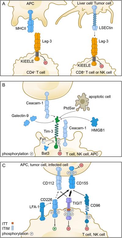
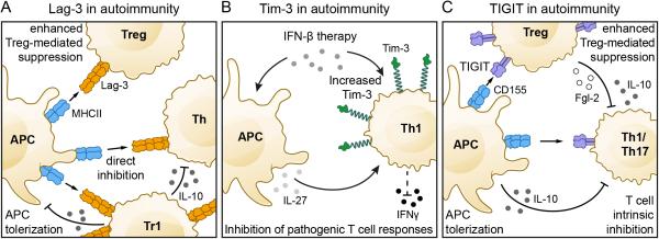
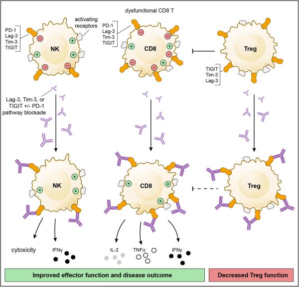
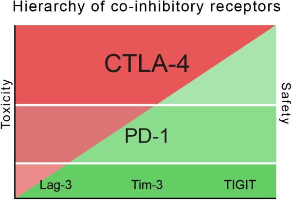
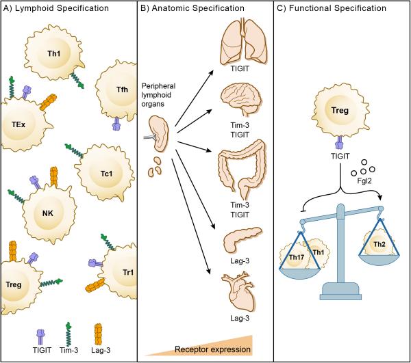
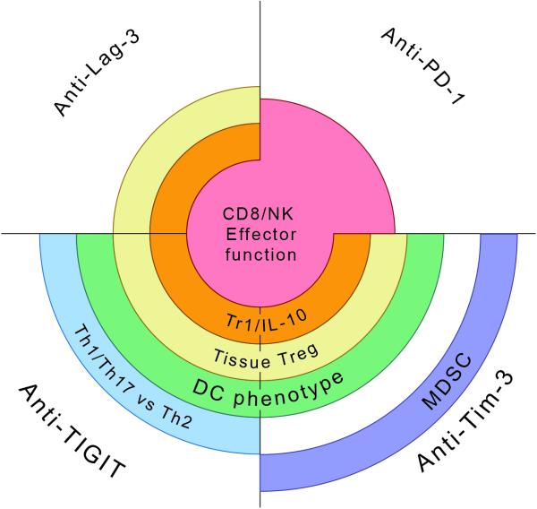
Co-inhibitory receptors, such as CTLA-4 and PD-1, have an important role in regulating T cell responses and have proven to be effective targets in the setting of chronic diseases where constitutive co-inhibitory receptor expression on T cells dampens effector T cell responses. Unfortunately, many patients still fail to respond to therapies that target CTLA-4 and PD-1. The next wave of co-inhibitory receptor targets that are being explored in clinical trials include Lag-3, Tim-3, and TIGIT. These receptors, although they belong to the same class of receptors as PD-1 and CTLA-4, exhibit unique functions, especially at tissue sites where they regulate distinct aspects of immunity. Increased understanding of the specialized functions of these receptors will inform the rational application of therapies that target these receptors to the clinic.


Figures

Figure 1
Figure 1. Co-inhibitory receptor pathways
A) The Lag-3 pathway. Left panel, Lag-3 is expressed on CD4+ T cells and binds to MHC class II on antigen presenting cells. Right panel, Lag-3 is expressed on CD8+ T cells and NK cells and binds to L-SECtin on tumor cells or liver cells. The cytoplasmic tail of Lag-3 contains a unique KIEELE motif that is essential for the inhibitory function of Lag-3. B) The Tim-3 pathway. Tim-3 is expressed on T cells, NK cells, and some APC. Tim-3 ligands include soluble ligands (galectin-9 and HMGB1) and cell surface ligands (Ceacam-1 and Phosphatidyl serine – PtdSer). Bat-3 and Fyn bind to the same region on the cytoplasmic tail of Tim-3. Ligand binding triggers the dissociation of Bat-3 from the cytoplasmic tail of Tim-3, thus allowing Fyn to bind and promote the inhibitory function of Tim-3. C) The CD226/TIGIT Pathway. CD226, TIGIT, and CD96 are expressed on T cells and NK cells and share the ligands CD112 and CD155, which are expressed on APCs and other cells such as tumor cells. CD226 associates with the integrin LFA-1 and delivers a positive signal. TIGIT, CD96, and CD155 contain ITIM motifs in their cytoplasmic tails and can deliver inhibitory signals. TIGIT further contains an ITT-like motif. CD155 and TIGIT exist as homodimers on the cell surface, and dimerization is essential for their proper function.
Figure 2
Figure 2. The Lag-3, Tim-3, and TIGIT pathways in autoimmunity
A) Lag-3 plays a protective role in autoimmunity by dampening T helper (Th) cell responses directly through engagement of MHCII. In addition, Lag-3 indirectly inhibits effector T cell responses via promotion of Treg and Tr1-mediated suppression. B) In autoimmune diseases such as MS, Tim-3 is under-expressed on pathogenic Th1 cells. IFN-β therapy can increase Tim-3 on antigen specific T cells directly or indirectly via promotion of IL-27 production from local antigen presenting cells. Increased expression of Tim-3 is associated with reduction in disease relapses. C) TIGIT inhibits auto-pathogenic Th1/Th17 T cell responses through three different pathways: 1) TIGIT directly inhibits T cell activation and expansion; 2) TIGIT expressing effector and regulatory T cells engage CD155 on APCs thereby inducing tolerogenic APCs that secrete IL-10; 3) TIGIT promotes Treg-mediated suppression through the induction of IL-10 and Fgl2, which potently and selectively suppress Th1 and Th17 responses.
Figure 3
Figure 3. The Lag-3, Tim-3, and TIGIT pathways in chronic diseases
Lag-3, Tim-3, and TIGIT are highly expressed on dysfunctional or exhausted T cells in chronic diseases such as chronic viral infection and cancer. In these diseases, combinatorial receptor blockade has strong synergistic effects, resulting in improved effector CD8+ T cell and NK cell function as well as decreased Treg-mediated suppression. These combined actions improve disease outcome.
Figure 4
Figure 4. Hierarchy of co-inhibitory receptors
Co-inhibitory receptors are ranked from top to bottom according to their impact on the maintenance of self-tolerance. The impact of a given co-inhibitory receptor on self-tolerance is directly proportional to the amount of autoimmune toxicity observed when the receptor is deficient either as a result of genetic loss or therapeutic modulation. Genetic and/or therapeutic modulation of co-inhibitory receptors at the top of the hierarchy (Tier 1) is predicted to be associated with more autoimmune-like toxicity than modulation of co-inhibitory receptors at the bottom of the hierarchy (Tier 2). Accordingly, Tier 2 co-inhibitory receptors are predicted to have a better safety profile in the clinic.
Figure 5
Figure 5. Specification of checkpoint-receptor pathways
A) Lymphoid specification. Some co-inhibitory receptors are preferentially expressed on distinct lymphocyte subsets. B) Anatomic specification. Some co-inhibitory receptor pathways may dominate in different tissue sites where ligands and/or receptors are highly expressed. C) Functional specification. Some co-inhibitory receptors may regulate distinct aspects of immunity such as the regulation of the balance between Type 1/Type 17 immunity and Type 2 immunity by TIGIT.
Figure 6
Figure 6. Immunological effects of checkpoint receptor blockades
Schematic representation showing the effects of PD-1, Lag-3, Tim-3, and TIGIT blockades on the immune response. While all checkpoint receptor blockades have some effect on CD8+ T cell and NK cell effector function, the effect of PD-1 blockade is proportionally larger than that of Lag-3, Tim-3, or TIGIT blockade alone. Lag-3, Tim-3, and TIGIT blockades will preferentially affect tumor tissue Treg and IL-10 producing Tr1 cells. Tim-3 and TIGIT blockades will additionally affect DC phenotype. A unique effect of TIGIT blockade is shifting the balance in favor of Type 1/17 immunity versus Type 2 immunity while a unique effect of Tim-3 blockade is to dampen MDSC. Thus, different checkpoint receptor blockades can be combined to achieve distinct effects on the immune response.

References

    1. Awasthi A, Carrier Y, Peron JP, Bettelli E, Kamanaka M, Flavell RA, Kuchroo VK, Oukka M, Weiner HL. A dominant function for interleukin 27 in generating interleukin 10-producing anti-inflammatory T cells. Nat Immunol. 2007;8:1380–1389. - PubMed
    1. Bettini M, Szymczak-Workman AL, Forbes K, Castellaw AH, Selby M, Pan X, Drake CG, Korman AJ, Vignali DA. Cutting edge: accelerated autoimmune diabetes in the absence of LAG-3. Journal of immunology. 2011;187:3493–3498. - PMC - PubMed
    1. Blackburn SD, Shin H, Haining WN, Zou T, Workman CJ, Polley A, Betts MR, Freeman GJ, Vignali DA, Wherry EJ. Coregulation of CD8+ T cell exhaustion by multiple inhibitory receptors during chronic viral infection. Nat Immunol. 2009;10:29–37. - PMC - PubMed
    1. Boivin N, Baillargeon J, Doss PM, Roy AP, Rangachari M. Interferon-beta suppresses murine Th1 cell function in the absence of antigen-presenting cells. PloS one. 2015;10:e0124802. - PMC - PubMed
    1. Boles KS, Vermi W, Facchetti F, Fuchs A, Wilson TJ, Diacovo TG, Cella M, Colonna M. A novel molecular interaction for the adhesion of follicular CD4 T cells to follicular DC. European journal of immunology. 2009;39:695–703. - PMC - PubMed

Publication types

MeSH terms