Review
. 2016 May 17;44(5):1034-51.
doi: 10.1016/j.immuni.2016.04.017.

Co-stimulatory and Co-inhibitory Pathways in Autoimmunity

Affiliations
Review

Co-stimulatory and Co-inhibitory Pathways in Autoimmunity

Qianxia Zhang et al. Immunity. .

Abstract

The immune system is guided by a series of checks and balances, a major component of which is a large array of co-stimulatory and co-inhibitory pathways that modulate the host response. Although co-stimulation is essential for boosting and shaping the initial response following signaling through the antigen receptor, inhibitory pathways are also critical for modulating the immune response. Excessive co-stimulation and/or insufficient co-inhibition can lead to a breakdown of self-tolerance and thus to autoimmunity. In this review, we will focus on the role of co-stimulatory and co-inhibitory pathways in two systemic (systemic lupus erythematosus and rheumatoid arthritis) and two organ-specific (multiple sclerosis and type 1 diabetes) emblematic autoimmune diseases. We will also discuss how mechanistic analysis of these pathways has led to the identification of potential therapeutic targets and initiation of clinical trials for autoimmune diseases, as well as outline some of the challenges that lie ahead.


Figures

Figure 1
Figure 1. Co-stimulatory and co-inhibitory pathways in Systemic Lupus Erythematosus (SLE)
The hierarchical utilization of co-stimulatory and co-inhibitory receptors and their corresponding ligands is compared in SLE. The extent to which a co-stimulatory or co-inhibitory molecule is utilized is indicted by dark (dominant utilization - red or blue) or light shading (partial utilization - pink or cyan). Molecules that appear to exhibit differential or controversial function (mixed bluered) in certain diseases, or for which there are no data (grey shading) are also noted.
Figure 2
Figure 2. Co-stimulatory and co-inhibitory pathways in Rheumatoid Arthritis (RA)
The hierarchical utilization of co-stimulatory and co-inhibitory receptors and their corresponding ligands is compared in RA. The extent to which a co-stimulatory or co-inhibitory molecule is utilized is indicted by dark (dominant utilization - red or blue) or light shading (partial utilization - pink or cyan). Molecules that appear to exhibit differential or controversial function (mixed bluered) in certain diseases, or for which there are no data (grey shading) are also noted.
Figure 3
Figure 3. Co-stimulatory and co-inhibitory pathways in Multiple Sclerosis (MS)
The hierarchical utilization of co-stimulatory and co-inhibitory receptors and their corresponding ligands is compared in MS. The extent to which a co-stimulatory or co-inhibitory molecule is utilized is indicted by dark (dominant utilization - red or blue) or light shading (partial utilization - pink or cyan). Molecules that appear to exhibit differential or controversial function (mixed bluered) in certain diseases, or for which there are no data (grey shading) are also noted.
Figure 4
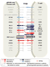
Figure 4. Co-stimulatory and co-inhibitory pathways in Type 1 Diabetes (T1D)
The hierarchical utilization of co-stimulatory and co-inhibitory receptors and their corresponding ligands is compared in T1D. The extent to which a co-stimulatory or co-inhibitory molecule is utilized is indicted by dark (dominant utilization - red or blue) or light shading (partial utilization - pink or cyan). Molecules that appear to exhibit differential or controversial function (mixed bluered) in certain diseases, or for which there are no data (grey shading) are also noted.

Similar articles

Cited by

References

    1. Amrani A, Serra P, Yamanouchi J, Han B, Thiessen S, Verdaguer J, Santamaria P. CD154-dependent priming of diabetogenic CD4(+) T cells dissociated from activation of antigen-presenting cells. Immunity. 2002;16:719–732. - PubMed
    1. Ansari MJ, Fiorina P, Dada S, Guleria I, Ueno T, Yuan X, Trikudanathan S, Smith RN, Freeman G, Sayegh MH. Role of ICOS pathway in autoimmune and alloimmune responses in NOD mice. Clin Immunol. 2008;126:140–147. - PubMed
    1. Ansari MJ, Salama AD, Chitnis T, Smith RN, Yagita H, Akiba H, Yamazaki T, Azuma M, Iwai H, Khoury SJ, et al. The programmed death-1 (PD-1) pathway regulates autoimmune diabetes in nonobese diabetic (NOD) mice. J Exp Med. 2003;198:63–69. - PMC - PubMed
    1. Araki M, Chung D, Liu S, Rainbow DB, Chamberlain G, Garner V, Hunter KM, Vijayakrishnan L, Peterson LB, Oukka M, et al. Genetic evidence that the differential expression of the ligand-independent isoform of CTLA-4 is the molecular basis of the Idd5.1 type 1 diabetes region in nonobese diabetic mice. J Immunol. 2009;183:5146–5157. - PMC - PubMed
    1. Atkinson MA, Eisenbarth GS, Michels AW. Type 1 diabetes. Lancet. 2014;383:69–82. - PMC - PubMed

Publication types

MeSH terms

Substances