Review
. 2017 Jan;8(1):10.1002/wrna.1366.
doi: 10.1002/wrna.1366. Epub 2016 May 18.

Ribosome-based quality control of mRNA and nascent peptides

Affiliations
Review

Ribosome-based quality control of mRNA and nascent peptides

Carrie L Simms et al. Wiley Interdiscip Rev RNA. 2017 Jan.

Abstract

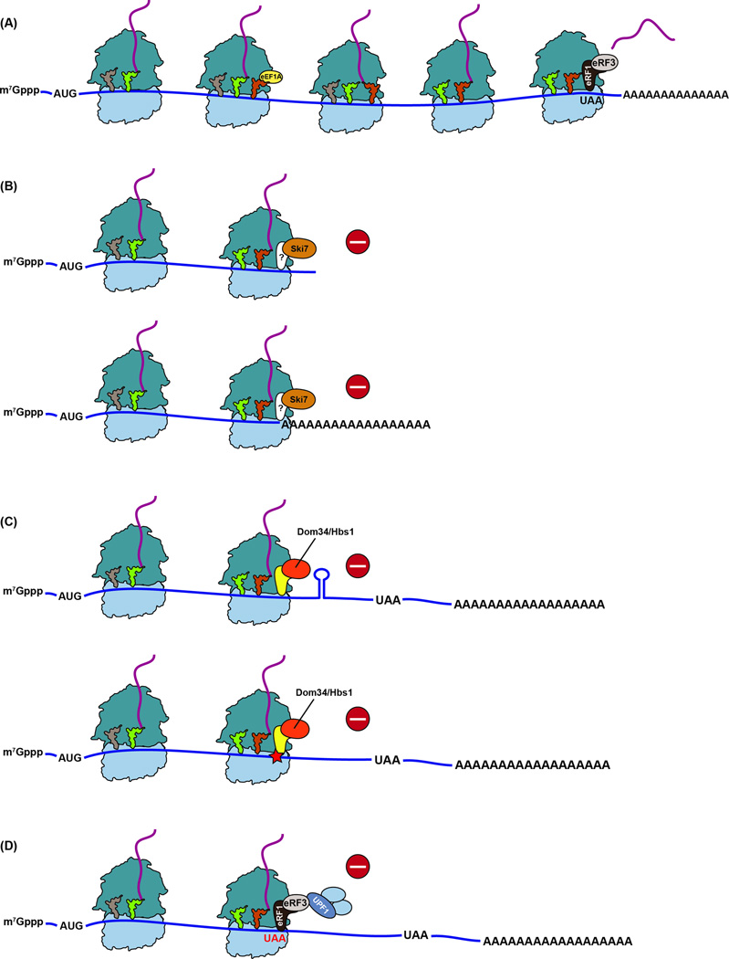
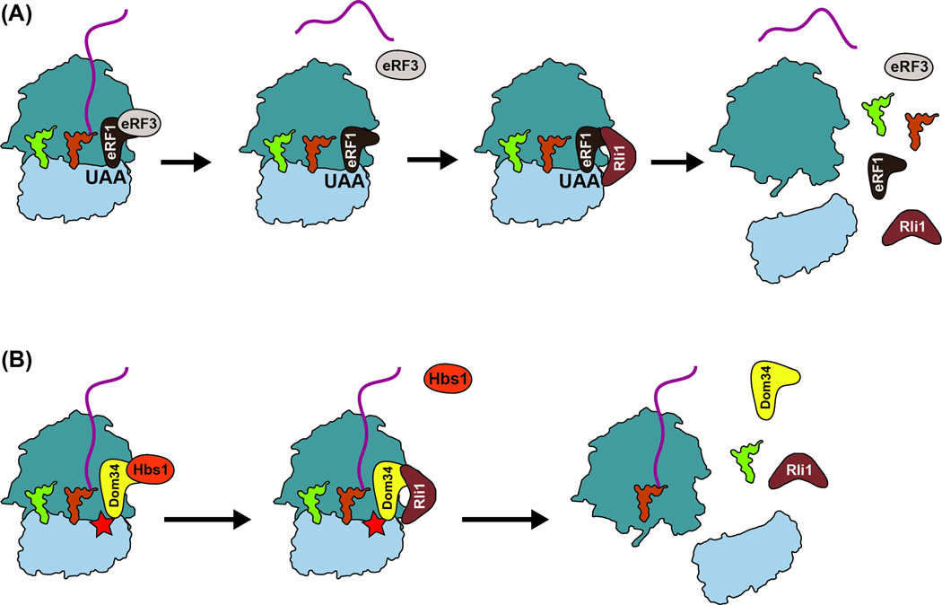
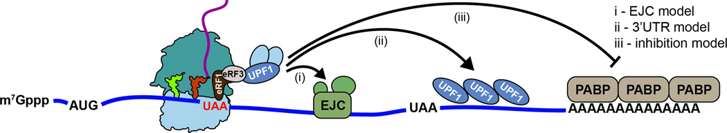
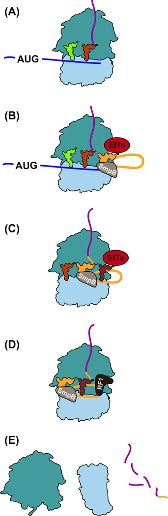
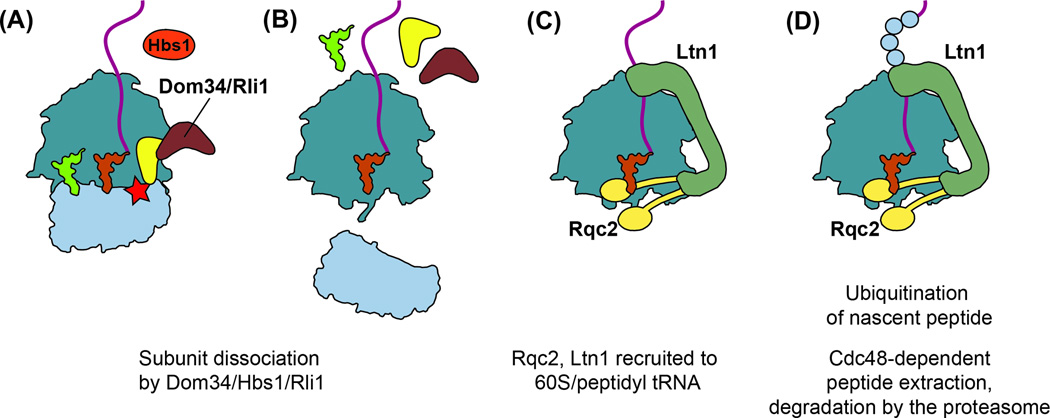
Quality control processes are widespread and play essential roles in detecting defective molecules and removing them in order to maintain organismal fitness. Aberrant messenger RNA (mRNA) molecules, unless properly managed, pose a significant hurdle to cellular proteostasis. Often mRNAs harbor premature stop codons, possess structures that present a block to the translational machinery, or lack stop codons entirely. In eukaryotes, the three cytoplasmic mRNA-surveillance processes, nonsense-mediated decay (NMD), no-go decay (NGD), and nonstop decay (NSD), evolved to cope with these aberrant mRNAs, respectively. Nonstop mRNAs and mRNAs that inhibit translation elongation are especially problematic as they sequester valuable ribosomes from the translating ribosome pool. As a result, in addition to RNA degradation, NSD and NGD are intimately coupled to ribosome rescue in all domains of life. Furthermore, protein products produced from all three classes of defective mRNAs are more likely to malfunction. It is not surprising then that these truncated nascent protein products are subject to degradation. Over the past few years, many studies have begun to document a central role for the ribosome in initiating the RNA and protein quality control processes. The ribosome appears to be responsible for recognizing the target mRNAs as well as for recruiting the factors required to carry out the processes of ribosome rescue and nascent protein decay. WIREs RNA 2017, 8:e1366. doi: 10.1002/wrna.1366 For further resources related to this article, please visit the WIREs website.


Figures

Figure 1
Figure 1. Translation of intact mRNAs versus aberrant mRNAs
(A) During normal translation, a ternary complex of aa-tRNA, eEF1A (EFTu in bacteria) and GTP binds the ribosome to decode the A site codon. Following peptidyl transfer, the elongation phase continues until a stop codon arrives at the A site, where it is recognized by eRF1 release factor in a complex with eRF3-GTP. Hydrolysis of the peptidyl tRNA and dissociation of eRF3 is triggered by conformational changes to eRF1 upon GTP hydrolysis. (B) In S. cerevisiae, the GTPase Ski7 interacts with the ribosome when it is stalled at the 3’ end of a stop-codon-less mRNA (top) or when it translates a polyA tail (bottom), activating non-stop decay (NSD). A binding partner for SKI7 has not been identified (shown as ?). (C) No-go decay (NGD) is responsible for recognizing and rescuing ribosomes stalled within an mRNA, either due to stable structures that block its progression (top) or caused by damaged nucleobases or strings of rare codons (bottom - shown as a star). Dom34, together with Hbs1-GTP, binds the ribosome and recycles the stalled ribosome. The process results in an endonucleolytic cleavage event (not shown), which may precede Dom34 recruitment. (D) Premature stop codons are recognized by canonical release factors that interact with Upf1 and other factors of the nonsense mediated decay (NMD) pathway.
Figure 2
Figure 2. Ribosome recycling
(A) During termination under normal conditions, eRF1/eRF3 release factors recognize a stop codon and bind the ribosome. GTP hydrolysis leads to conformational changes in eRF1, which mediates release of the peptide and recruitment of Rli1. Rli1 is required to promote ribosome splitting after hydrolysis of ATP. (B) During NGD, Dom34/Hbs1 recognize a stalled ribosome and bind the A site. Upon GTP hydrolysis, Hbs1 dissociates, allowing interaction with Rli1 and the subunits dissociate, but without release of the peptidyl tRNA.
Figure 3
Figure 3. Models for nonsense-mediated decay
mRNAs containing premature stop codons are recognized by the cell using several possible mechanisms. (i) the EJC model relies on interactions between an EJC located downstream of the premature stop codon and the Upf proteins that are bound to release factors on the ribosome. (ii) The 3’ UTR model suggests that Upf1 coats the UTR and the local concentration of the protein distinguishes NMD targets from other mRNAs. (iii) Interactions between eRF3 and polyA binding protein (PABP), essential during normal termination, are inhibited when the distance between the premature stop codon and polyA tail is large.
Figure 4
Figure 4. Ribosome rescue by trans-translation in bacteria
(A) Ribosomes stall at the 3’ end of non-stop mRNAs or those containing rare codons. (B) A complex consisting of tmRNA, SmpB and EF-Tu, together with GTP, binds to the A site. (C) The nascent peptide is transferred to tmRNA and translation resumes on the ssrA ORF, tagging the defective protein at its 3’ end. The mRNA is released and degraded. (D) Termination occurs on the tmRNA stop codon using standard release factors. (E) Ribosomes dissociate and the tagged protein is degraded.
Figure 5
Figure 5. Co-translational protein quality control
(A–B) Ribosomes stalled on defective mRNAs are released through the NGD pathway. (C) Rqc2 binds the 60S subunit, contacting the exposed P site tRNA and stabilizing Ltn1 binding. The C-terminal RING domain of Ltn1 contacts the exit tunnel while the N-terminus interacts with the sarcin-ricin loop on the ribosome. (D) Ltn1 ubiquitinates the nascent peptide, targeting it for degradation by the proteasome. Extraction of the peptide from the ribosome depends on Cdc48.

References

    1. Wolff S, Weissman JS, Dillin A. Differential Scales of Protein Quality Control. Cell. 2014;157:52–64. - PubMed
    1. Drummond DA, Wilke CO. Mistranslation-induced protein misfolding as a dominant constraint on coding-sequence evolution. Cell. 2008;134:341–352. - PMC - PubMed
    1. Simms CL, Hudson BH, Mosior JW, Rangwala AS, Zaher HS. An active role for the ribosome in determining the fate of oxidized mRNA. Cell Rep. 2014;9:1256–1264. - PMC - PubMed
    1. Wurtmann EJ, Wolin SL. RNA under attack: cellular handling of RNA damage. Crit Rev Biochem Mol Biol. 2009;44:34–49. - PMC - PubMed
    1. Ishigame H, Mosaheb MM, Sanjabi S, Flavell RA. Truncated Form of TGF-beta RII, But Not Its Absence, Induces Memory CD8(+) T Cell Expansion and Lymphoproliferative Disorder in Mice. Journal of Immunology. 2013;190:6340–6350. - PMC - PubMed

Publication types

LinkOut - more resources