Review
. 2016 May 23:11:77-100.
doi: 10.1146/annurev-pathol-012615-044231.

Hypoxia and Mucosal Inflammation

Affiliations
Review

Hypoxia and Mucosal Inflammation

Sean P Colgan et al. Annu Rev Pathol. .

Abstract

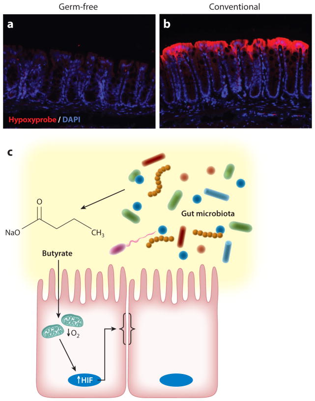
Sites of inflammation are defined by significant changes in metabolic activity. Recent studies have suggested that O2 metabolism and hypoxia play a prominent role in inflammation so-called "inflammatory hypoxia," which results from a combination of recruited inflammatory cells (e.g., neutrophils and monocytes), the local proliferation of multiple cell types, and the activation of multiple O2-consuming enzymes during inflammation. These shifts in energy supply and demand result in localized regions of hypoxia and have revealed the important function off the transcription factor HIF (hypoxia-inducible factor) in the regulation of key target genes that promote inflammatory resolution. Analysis of these pathways has provided multiple opportunities for understanding basic mechanisms of inflammation and has defined new targets for intervention. Here, we review recent work addressing tissue hypoxia and metabolic control of inflammation and immunity.

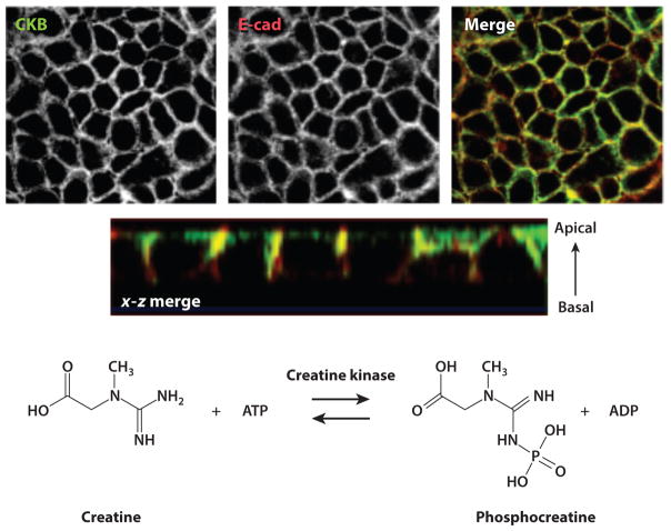
Keywords: colitis; creatine; epithelium; inflammation; metabolism; mucosa; murine model; neutrophil; nucleoside; nucleotidase; nucleotide; phosphocreatine.


Figures

Figure 1
Figure 1
Countercurrent blood flow and physiological hypoxia in the healthy intestinal mucosa. (a) A model of blood flow dynamics in the healthy intestinal mucosa. Countercurrent blood flow reduces local pO2 along the crypt-villus axis and results in low pO2 at the villus tip. (b) The mechanism of nitroimidazole dye retention in hypoxic regions. These imidazole derivatives (R-NO2) are taken into cells passively and reduced to highly reactive nitrogen intermediates ( R-NO2-·) within the cytoplasm. In the absence of adequate O2 to regenerate the native compound, these intermediates react with thiol groups in proteins, peptides, and amino acids to form adducts (R-NH2) where all atoms of the ring and side chain of the 2-nitroimidazole are retained at pO2 < 10 mm Hg. The adducts can be visualized through the use of labeled antibodies. (c) Physiological hypoxia. In the colonic mucosa of healthy mice, small amounts of nitroimidazole adduct (red ) are detected along the luminal aspect, suggesting a degree of physiological hypoxia in the normal colon. Abbreviation: pO2, partial oxygen pressure.
Figure 2
Figure 2
Role of PMN in HIF stabilization during mucosal inflammation. Tissue from the distal colons of healthy mice and mice with active TNBS colitis, with and without depletion of systemic PMN through the use of anti-GR-1 antibody, reveals an increase in HIF among colitic mice with PMN, which is attenuated in the absence of PMN. Staining for HIF ( green) and Ly6g (red ) indicates accumulation of PMN in TNBS colitis. DAPI stains (blue) signify areas of high DNA concentration. Scale bars = 100 μm. Abbreviations: HIF, hypoxia-inducible factor; PMN, polymorphonuclear leukocytes; TNBS, trinitrobenzene sulfonic.
Figure 3
Figure 3
Ulcerative colitis patients with crypt abscesses reveal hypoxia-dependent target induction. Biopsies from uninflamed margins (panels a and c) and inflamed regions with active crypt abscesses (panels b and d ) in patients with ulcerative colitis were processed for H&E (top) and stained (bottom) for hypoxia-responsive Glut-1 ( green) and neutrophil p47phox (red ). DAPI stains (blue) indicate nuclei. Scale bars = 50 μm. Abbreviation: H&E, hematoxylin and eosin. Figure adapted from Reference with permission.
Figure 4
Figure 4
O2-dependent regulation of AMPs; PMN fate in chronic inflammation and during resolution. (❶) Hypoxia: PMN induce localized hypoxia via NADPH oxidase activity, resulting in bacterial death by ROS generation. Hypoxia prolongs PMN life span, thereby preventing apoptosis. HIF-dependent AMPs are released and aid in antimicrobial activity. HIF-dependent glycolytic and proinflammatory genes (e.g., COX-2) are transcribed. RvE/RvD require the oxygenase activity of COX-2 and are synthesized. Activated PMN convert arachidonic acid to LXA4. (❷) Anoxia: In the absence of infection clearance, PMN accumulate and localized O2 depletion prevents NADPH oxidase activity. Under such conditions, PMN undergo NETosis or necrosis. (❶) Reoxygenation:With pathogenic bacterial clearance, PMN undergo apoptosis. RvE/RvD enhance clearance of apoptotic PMN through efferocytosis and induce intestinal ALPI expression on epithelia to restore mucosal homeostasis. Abbreviations: ALPI, alkaline phosphatase; AMP, antimicrobial peptide; HIF, hypoxia-inducible factor; PMN, polymorphonuclear leukocytes; ROS, reactive oxygen species; RvE/RvD, resolvins.
Figure 5
Figure 5
Creatine kinase expression at epithelial junctions. The images in the upper panels, obtained through confocal microscopy, show CKB (top left), the adherens junction marker E-cad (top middle), and two views of a colocalized (merged) stain (top right and second row). At the bottom is the enzymatic reaction catalyzed by creatine kinase. Abbreviations: ADP, adenosine diphosphate; ATP, adenosine triphosphate; CKB, creatine kinase, brain isoform; E-cad, E-cadherin. Figure adapted from Reference with permission.
Figure 6
Figure 6
Microbial-derived signals deplete local O2. The top panels depict hypoxia localization (red ), illuminated by the nitroimidazole dye pimonidazole, in (a) germ-free and (b) conventional animals. DAPI stains (blue) indicate areas of high DNA concentration. (c) A model in which microbial-derived short-chain fatty acids (e.g., butyrate) stimulate epithelial metabolism and deplete intracellular O2 to the extent that HIF-1α is stabilized and promotes epithelial barrier function. Abbreviation: HIF, hypoxia-inducible factor.

References

    1. Kominsky DJ, Campbell EL, Colgan SP. Metabolic shifts in immunity and inflammation. J Immunol. 2010;184:4062–68. - PMC - PubMed
    1. Colgan SP, Taylor CT. Hypoxia: an alarm signal during intestinal inflammation. Nat Rev Gastroenterol Hepatol. 2010;7:281–87. - PMC - PubMed
    1. Lewis JS, Lee JA, Underwood JC, Harris AL, Lewis CE. Macrophage responses to hypoxia: relevance to disease mechanisms. J Leukoc Biol. 1999;66:889–900. - PubMed
    1. Fox CJ, Hammerman PS, Thompson CB. Fuel feeds function: energy metabolism and the T-cell response. Nat Rev Immunol. 2005;5:844–52. - PubMed
    1. Sitkovsky M, Lukashev D. Regulation of immune cells by local-tissue oxygen tension: HIF1α and adenosine receptors. Nat Rev Immunol. 2005;5:712–21. - PubMed

LinkOut - more resources