

Review
doi: 10.1007/s00261-015-0589-3.
DCE MRI of prostate cancer
Affiliations
- PMID: 27193787
- PMCID: PMC6462146
- DOI: 10.1007/s00261-015-0589-3
Item in Clipboard
Review
DCE MRI of prostate cancer
Abdom Radiol (NY).
2016 May.
Abstract
DCE MRI is an established component of multi-parametric MRI of the prostate. The sequence highlights the vascularization of cancerous lesions, allowing readers to corroborate suspicious findings on T2W and DW MRI and to note subtle lesions not visible on the other sequences. In this article, we review the technical aspects, methods of evaluation, limitations, and future perspectives of DCE MRI.
Keywords: DCE MRI; Multi-parametric MRI; Prostate cancer.
Figures
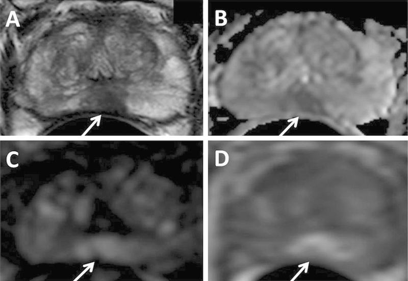
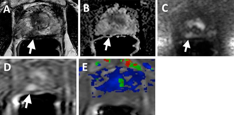
A 75-year-old man with PSA 10.23 ng/mL with no prior biopsy history. Axial T2W MRI (A) shows a hypointense lesion in the midline apical peripheral zone, which shows restricted diffusion on ADC map (B) and b2000 DW MRI (C) (arrows). The lesion shows positive enhancement on DCE MRI (D) (arrow). The lesion was found to have Gleason 8(4 + 4) prostate adenocarcinoma on targeted biopsy.
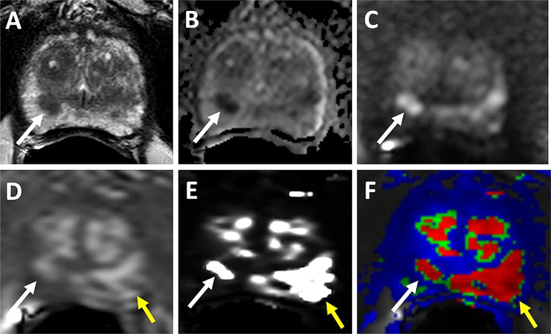
69-year-old man with PSA of 34.95 ng/mL. T2W (A), ADC map (B), b2000 DWI (C), raw DCE MRI (D) show a right mid peripheral zone lesion (Gleason 3 + 4 cancer at targeted biopsy). Raw DCE MRI (D), kep (E) and Ktrans (F) mps show an additional lesion in the left mid peripheral zone (yellow arrows), which has subtle positive features on T2W MRI and DW MRI. This lesion includes Gleason 4 + 5 prostate cancer.
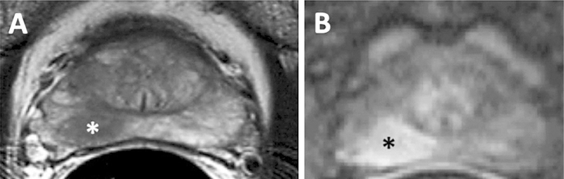
MRI with A T2 weighted and B DCE MRI in 67-year-old-man with a biopsy proven Gleason 3 + 4 in the right mid peripheral zone demonstrating an area of T2 hypointensity and an area of early enhancement (asterisks).
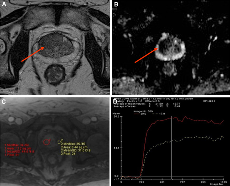
Multi-parametric MRI with A T2-weighted B ADC map C DCE and D time-intensity curve of the prostate on a 62-year-old man with a rising PSA of 10.8 and a previous negative prostate biopsy demonstrates a large area of low T2 signal intensity (arrow) with restricted diffusion with ADC of 877 (arrow) and hyperenhancement (red circle) demonstrating a Type 3 curve (red curve) compared normal tissue (yellow circle) demonstrating a Type 1 curve (white curve). Subsequent MRI-US fusion biopsy revealed Gleason 3 + 4.
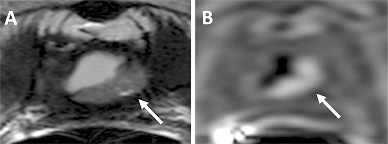
68-year-old man with a PSA = 11 ng/mL 6 years after prostatectomy. T2W MRI shows a lesion in the prostatectomy bed (arrow) (A), DCE MRI shows hyperenhancement within the lesion confirming local recurrence (arrow) (B).
70-year-old man with a PSA of 5.67 ng/mL. T2W MRI (A), ADC map (B), b2000 DWI (C), DCE MRI (D) shows a right mid peripheral zone lesion (arrows). Ktrans map derived from DCE MRI (E) is obscured due to motion artifacts and lesion is not localized in the map (supplementary video).
References
-
- Humphrey PA (2014) Cancers of the male reproductive organs. In: Stewart BW, Wild CP (eds) World cancer report Lyon: World Health Organization
-
- Delongchamps NB, Singh A, Haas GP (2006) The role of prevalence in the diagnosis of prostate cancer. Cancer Control 13(3):158–168 - PubMed
-
- Ries LAG, Melbert D, Krapcho M, et al. (eds) (1975–2004) SEER cancer statistics review, National Cancer Institute, Bethesda, MD: 2007. Available on http://seer.cancer.gov/csr/1975_2004/
-
- Fütterer JJ, Briganti A, De Visschere P, et al. (2015) Can clinically significant prostate cancer be detected with multiparametric magnetic resonance imaging? A systematic review of the literature. Eur Urol 68(6):1045–1053 - PubMed
Publication types
MeSH terms
Substances
Grants and funding
LinkOut - more resources
Full Text Sources
Other Literature Sources
Medical
