

Visualizing and Analyzing Branching Microtubule Nucleation Using Meiotic Xenopus Egg Extracts and TIRF Microscopy
- PMID: 27193844
- PMCID: PMC5016078
- DOI: 10.1007/978-1-4939-3542-0_6
Visualizing and Analyzing Branching Microtubule Nucleation Using Meiotic Xenopus Egg Extracts and TIRF Microscopy
Abstract
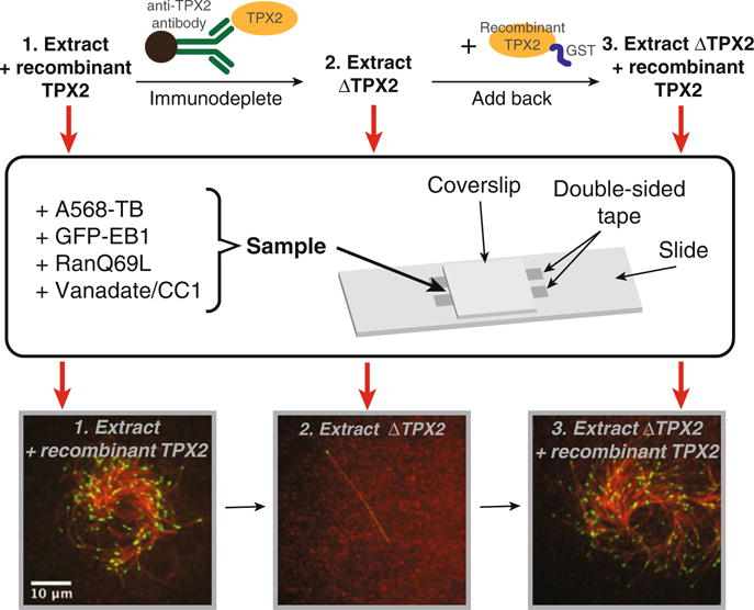
Mitotic and meiotic spindles consist primarily of microtubules, which originate from centrosomes and within the vicinity of chromatin. Indirect evidence suggested that microtubules also originate throughout the spindle, but the high microtubule density within the spindle precludes the direct observation of this phenomenon. By using meiotic Xenopus laevis egg extract and employing total internal reflection (TIRF) microscopy, microtubule nucleation from preexisting microtubules could be demonstrated and analyzed. Branching microtubule nucleation is an ideal mechanism to assemble and maintain a mitotic spindle, because microtubule numbers are amplified while preserving their polarity. Here, we describe the assays that made these findings possible and the experiments that helped identify the key molecular players involved.
Keywords: Cell division; Cytoskeleton; Meiotic spindle; Microtubule; Microtubule nucleation; Mitotic spindle; TIRF microscopy; Xenopus laevis egg extract.
Figures
Similar articles
-
Dissecting Protein Complexes in Branching Microtubule Nucleation Using Meiotic Xenopus Egg Extracts.Cold Spring Harb Protoc. 2018 Sep 4;2018(9):pdb.prot100958. doi: 10.1101/pdb.prot100958. Cold Spring Harb Protoc. 2018. PMID: 29321281 Free PMC article.
-
Branching microtubule nucleation in Xenopus egg extracts mediated by augmin and TPX2.Cell. 2013 Feb 14;152(4):768-77. doi: 10.1016/j.cell.2012.12.044. Cell. 2013. PMID: 23415226 Free PMC article.
-
Autocatalytic microtubule nucleation determines the size and mass of Xenopus laevis egg extract spindles.Elife. 2018 Jan 11;7:e31149. doi: 10.7554/eLife.31149. Elife. 2018. PMID: 29323637 Free PMC article.
-
The chromosomal basis of meiotic acentrosomal spindle assembly and function in oocytes.Chromosoma. 2017 Jun;126(3):351-364. doi: 10.1007/s00412-016-0618-1. Epub 2016 Nov 11. Chromosoma. 2017. PMID: 27837282 Free PMC article. Review.
-
Microtubule dynamics in Xenopus egg extracts.Microsc Res Tech. 1999 Mar 15;44(6):435-45. doi: 10.1002/(SICI)1097-0029(19990315)44:6<435::AID-JEMT5>3.0.CO;2-T. Microsc Res Tech. 1999. PMID: 10211677 Review.
Cited by
-
A hydrodynamic instability drives protein droplet formation on microtubules to nucleate branches.Nat Phys. 2021 Apr;17(4):493-498. doi: 10.1038/s41567-020-01141-8. Epub 2021 Jan 28. Nat Phys. 2021. PMID: 35211183 Free PMC article. No abstract available.
-
Motorless transport of microtubules along tubulin, RanGTP, and salt gradients.Nat Commun. 2024 Nov 1;15(1):9434. doi: 10.1038/s41467-024-53656-w. Nat Commun. 2024. PMID: 39487112 Free PMC article.
-
Acentrosomal spindles assemble from branching microtubule nucleation near chromosomes in Xenopus laevis egg extract.Nat Commun. 2023 Jun 21;14(1):3696. doi: 10.1038/s41467-023-39041-z. Nat Commun. 2023. PMID: 37344488 Free PMC article.
-
Protein interactomes of protein phosphatase 2A B55 regulatory subunits reveal B55-mediated regulation of replication protein A under replication stress.Sci Rep. 2018 Feb 8;8(1):2683. doi: 10.1038/s41598-018-21040-6. Sci Rep. 2018. PMID: 29422626 Free PMC article.
-
Mechanism of how augmin directly targets the γ-tubulin ring complex to microtubules.J Cell Biol. 2018 Jul 2;217(7):2417-2428. doi: 10.1083/jcb.201711090. Epub 2018 Jun 6. J Cell Biol. 2018. PMID: 29875259 Free PMC article.
References
-
- Lajoie-Mazenc I, Tollon Y, Detraves C, Julian M, Moisand A, Gueth-Hallonet C, Debec A, Salles-Passador I, Puget A, Mazarguil H, et al. Recruitment of antigenic gamma-tubulin during mitosis in animal cells: presence of gamma-tubulin in the mitotic spindle. J Cell Sci. 1994;107(Pt 10):2825–2837. - PubMed
-
- Mahoney NM, Goshima G, Douglass AD, Vale RD. Making microtubule sand mitotic spindles in cells without functional centrosomes. Curr Biol. 2006;16:564–569. - PubMed
-
- Brugues J, Nuzzo V, Mazur E, Needleman DJ. Nucleation and transport organize microtubules in metaphase spindles. Cell. 2012;149:554–564. - PubMed
Publication types
MeSH terms
Substances
Grants and funding
LinkOut - more resources
Full Text Sources
Other Literature Sources
