. 2016 Aug;54(8):439-46.
doi: 10.1002/dvg.22949. Epub 2016 Jun 3.

Cre-dependent DREADD (Designer Receptors Exclusively Activated by Designer Drugs) mice

Affiliations

Cre-dependent DREADD (Designer Receptors Exclusively Activated by Designer Drugs) mice

Hu Zhu et al. Genesis. 2016 Aug.

Abstract

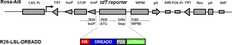
DREADDs, designer receptors exclusively activated by designer drugs, are engineered G protein-coupled receptors (GPCR) which can precisely control GPCR signaling pathways (for example, Gq, Gs, and Gi). This chemogenetic technology for control of GPCR signaling has been successfully applied in a variety of in vivo studies, including in mice, to remotely control GPCR signaling, for example, in neurons, glia cells, pancreatic β-cells, or cancer cells. In order to fully explore the in vivo applications of the DREADD technology, we generated hM3Dq and hM4Di strains of mice which allow for Cre recombinase-mediated restricted expression of these pathway-selective DREADDs. With the many Cre driver lines now available, these DREADD lines will be applicable to studying a wide array of research and preclinical questions. genesis 54:439-446, 2016. © 2016 Wiley Periodicals, Inc.

Keywords: G protein-coupled receptors; GsD; chemogenetic; hM3Dq; hM4Di.


Figures

FIG. 1
FIG. 1
DREADD conditional targeting vectors. Schematic of Ai9 targeting vector (drawing and sequence annotations after Madison et al., 2010). Replacement of the tdTomato sequence by HA-DREADD-P2A-mCitrine generated R26-LSL-DREADD vectors.
FIG. 2
FIG. 2
ES cells express DREAADs after Cre-mediated removal of STOP. Colonies from R26-LSL-hM4Di-DREADD-expressing ES cells were fluorescent only when transformed with CMV-Cre (A, B), while non-Cre transfected cells showed no fluorescence (C, D). To visualize membrane localization of DREADDs at the single cell level, Cre recombinase transformed ES cells were plated directly on gelatinized coverslips at low density; cells plated under these conditions show a flat, fibroblast-like morphology. Cells were fixed 2 days later, and treated with primary and secondary antibodies to HA. Confocal microscopy allowed visualizing, separately, cytoplasm and membrane of the same cells. Citrine (green) localizes to the cytoplasm (E), while HA (red) is fused to DREADDs and localizes to the cell membrane (F).
FIG. 3
FIG. 3
Conditional DREAAD mice express ubiquitously after CMV-Cre-mediated removal of STOP. R26-LSL-hM4Di mice were crossed with CMV-Cre driver mice. Various organs from offspring were collected, fixed, and sectioned. Shown are sections of lung (A), heart (B), tail (C), and ear (D) imaged under fluorescent light. Solid scale bar, 100 μm; dashed scale bar, 50 μm.
FIG. 4
FIG. 4
Conditional DREAAD mice express in targeted brain regions after CamKIIa-Cre-mediated removal of STOP. R26-LSL-hM4Di- and –hM3Dq-DREADD mice were crossed with CamKIIa-Cre driver mice. Coronal sections of offspring (A – hM4Di, B – hM3Dq) were stained with anti-GFP antibody and imaged (20×). (A) Dentate gyrus of the hippocampus shows granular cells fluorescent. (B) Cerebral cortex shows pyramidal cells fluorescent. Scale bar: 100 μm.
FIG. 5
FIG. 5
Conditional DREAAD expression in targeted brain regions after Emx1-Cre-mediated removal of STOP. R26-LSL-hM4Di- and –hM3Dq-DREADD mice were crossed with Emx1-Cre driver mice. (A, B) Coronal sections of offspring were stained for mCitrine (GFP antibody) or HA. Tiled images of coronal sections of Emx1-Cre+/hM3Dq+ and Emx1-Cre-/hM3Dq+ (A) and of Emx1-Cre+/hM4Di+ and Emx1-Cre-/hM4Di+ (B) showing no transgene expression in the absence of Cre-recombinase. (C) Confocal images of neurons expressing HA-tagged hM3Dq at the membrane of mCitrine positive neurons in the presence of Cre-recombinase. (D) 10× tiled-image of an Emx1-Cre+/hM4Di+ mouse (white arrow indicating the mossy-fiber projections to CA3 from the dentate gyrus) and confocal images of HA-tagged hM4Di at the membrane of mCitrine positive CA3 targeting axons in the presence of Cre-recombinase.
FIG. 6
FIG. 6
Conditional DREADD mice display behavioral changes after clozapine-n-oxide (CNO) injection. R26-LSL-hM3Dq or R26-LSL-hM4Di mice were crossed with Emx1-Cre driver mice to obtain offspring with DREADD expression (EMX-hM3Dq or EMX-hM4Di) and respective control littermates without DREADD expression. In the open field test (A–D), the EMX-hM3Dq mice (circles) show significantly increased ambulation starting from 20 and lasting to 40 minutes after CNO injection (0.2 mg/kg, closed circles, n = 7) compared to vehicle injection (open circles, n = 7) (A, B). The control mice without hM3Dq display no difference in ambulation with CNO (closed triangles, n = 7) compared to vehicle (open triangles, n = 7) (C, D). In a fear conditioning test (E–H), the EMX-hM4Di mice showed significantly disrupted contextual memory compared to the control littermates. Mice were conditioned on day 1 and vehicle or CNO (2 mg/kg) administed in three subsequent injections at 0, 2 and 4 hours of conditioning. Freezing response to conditioning was similar in hM4Di mice (circles) and control mice (triangles) in both CNO (n = 10, each; filled symbols) (E) or vehcle (n = 8, each; open symbols) (F) treatment groups (CS, conditioned stimulus; US, unconditioned stimulus). Evaluation of contexual memory after 24 hours revealed that CNO treatment significantly disrupted contexual learning (p < 0.05) in the EMX-hM4Di mice (grey bars) than in the control mice (white bars) (G). The cued memory tested after 48 hours show no difference in cued learning between the EMX-hM4Di or the control mice (H). Data were expressed as Mean ± S.E.

References

    1. Alexander GM, Rogan SC, Abbas AI, Armbruster BN, Pei Y, Allen JA, Nonneman RJ, Hartmann J, Moy SS, Nicolelis MA, McNamara JO, Roth BL. Remote Control of Neuronal Activity in Transgenic Mice Expressing Evolved G Protein-Coupled Receptors. Neuron. 2009;63:27–39. - PMC - PubMed
    1. Armbruster BN, Li X, Pausch MH, Herlitze S, Roth BL. Evolving the Lock to Fit the Key to Create a Family of G Protein-Coupled Receptors Potently Activated by an Inert Ligand. Proc Natl Acad Sci USA. 2007;104:5163–68. - PMC - PubMed
    1. Gorski JA, Talley T, Qiu M, Puelles L, Rubenstein JLR, Jones KR. Cortical Excitatory Neurons and Glia, but Not GABAergic Neurons, Are Produced in the Emx1-Expressing Lineage. J Neurosci. 2002;22:6309–14. - PMC - PubMed
    1. Griesbeck O, Baird GS, Campbell RE, Zacharias DA, Tsien RY. Reducing the Environmental Sensitivity of Yellow Fluorescent Protein. Mechanism and Applications. J Biol Chem. 2001;276:29188–94. - PubMed
    1. Kim JH, Lee SR, Li LH, Park HJ, Park JH, Lee KY, Kim MK, Shin BA, Choi SY. High Cleavage Efficiency of a 2A Peptide Derived from Porcine Teschovirus-1 in Human Cell Lines, Zebrafish and Mice. PloS One. 2011;6:e18556. - PMC - PubMed

Publication types

LinkOut - more resources