. 2016 Sep;229(3):436-49.
doi: 10.1111/joa.12486. Epub 2016 May 19.

Cardiac troponin T is necessary for normal development in the embryonic chick heart

Affiliations

Cardiac troponin T is necessary for normal development in the embryonic chick heart

Jennifer England et al. J Anat. 2016 Sep.

Abstract

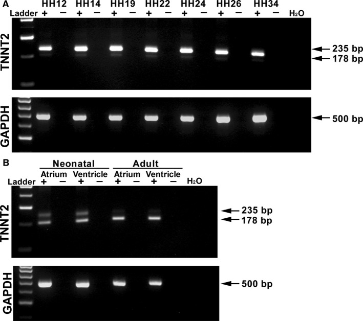
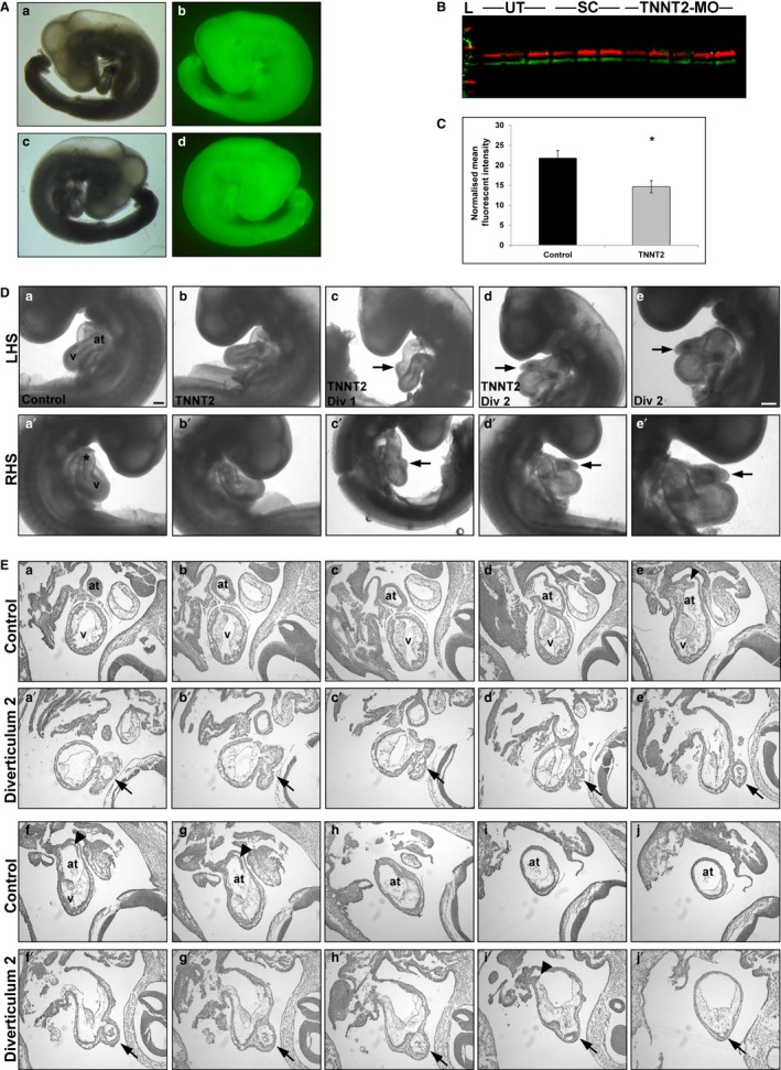
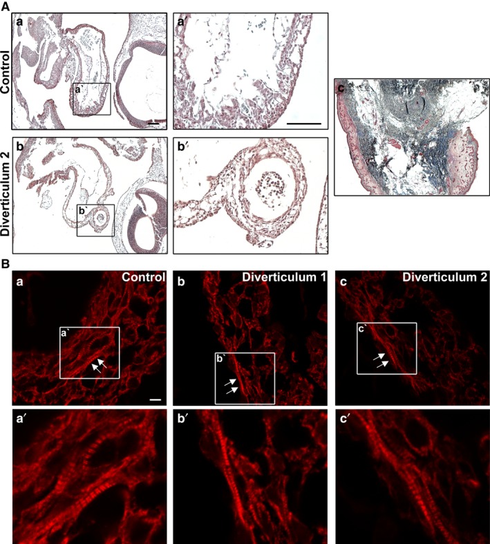
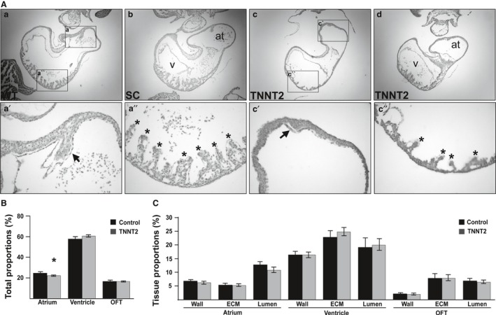
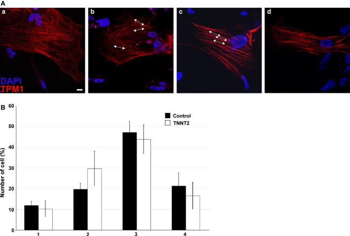
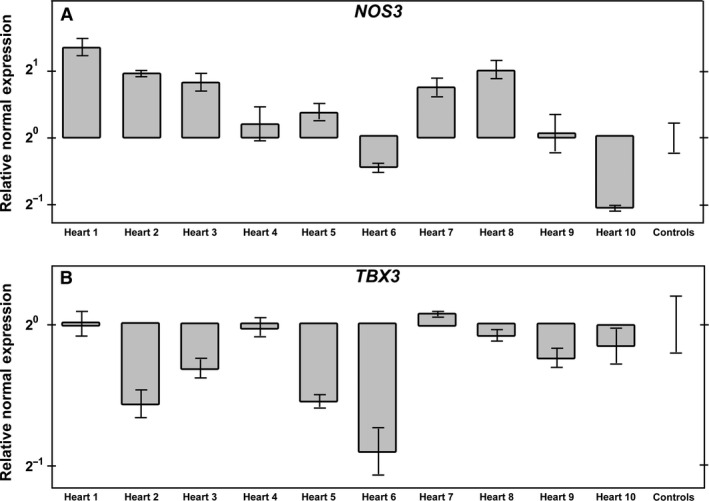
The heart is the first functioning organ to develop during embryogenesis. The formation of the heart is a tightly regulated and complex process, and alterations to its development can result in congenital heart defects. Mutations in sarcomeric proteins, such as alpha myosin heavy chain and cardiac alpha actin, have now been associated with congenital heart defects in humans, often with atrial septal defects. However, cardiac troponin T (cTNT encoded by gene TNNT2) has not. Using gene-specific antisense oligonucleotides, we have investigated the role of cTNT in chick cardiogenesis. TNNT2 is expressed throughout heart development and in the postnatal heart. TNNT2-morpholino treatment resulted in abnormal atrial septal growth and a reduction in the number of trabeculae in the developing primitive ventricular chamber. External analysis revealed the development of diverticula from the ventricular myocardial wall which showed no evidence of fibrosis and still retained a myocardial phenotype. Sarcomeric assembly appeared normal in these treated hearts. In humans, congenital ventricular diverticulum is a rare condition, which has not yet been genetically associated. However, abnormal haemodynamics is known to cause structural defects in the heart. Further, structural defects, including atrial septal defects and congenital diverticula, have previously been associated with conduction anomalies. Therefore, to provide mechanistic insights into the effect that cTNT knockdown has on the developing heart, quantitative PCR was performed to determine the expression of the shear stress responsive gene NOS3 and the conduction gene TBX3. Both genes were differentially expressed compared to controls. Therefore, a reduction in cTNT in the developing heart results in abnormal atrial septal formation and aberrant ventricular morphogenesis. We hypothesize that alterations to the haemodynamics, indicated by differential NOS3 expression, causes these abnormalities in growth in cTNT knockdown hearts. In addition, the muscular diverticula reported here suggest a novel role for mutations of structural sarcomeric proteins in the pathogenesis of congenital cardiac diverticula. From these studies, we suggest TNNT2 is a gene worthy of screening for those with a congenital heart defect, particularly atrial septal defects and ventricular diverticula.

Keywords: atrial septal defects; cardiac troponin T; congenital cardiac diverticula; heart development; structural protein.


Figures

Figure 1
Figure 1
mRNA expression of TNNT2 in the embryonic, neonatal and adult chick heart. PCR products obtained using a primer pair designed around the exon 5, which is alternately spliced in TNNT2. (A) Expression of a TNNT2 isoform in the embryonic chick heart (HH12–34; 235 bp). A smaller band of 178 bp can also be seen faintly expressed. GAPDH was used as a loading control (500 bp). (B) Two bands can be clearly seen in both the atrium and ventricle of the neonatal heart. In the adult atrium and ventricle, however, the lower band has a stronger signal than the upper 235 bp band. GAPDH was used as a loading control. + indicates RT; −, noRT; bp, base pairs; H2O, PCR control.
Figure 2
Figure 2
TNNT2‐MO treatment results in a knockdown of cTNT and external analysis reveals diverticula protruding from the heart wall. (A) Embryos were treated at HH11 with a TNNT2 or standard control fluorescein‐tagged morpholino; only embryos showing a strong fluorescent signal were harvested (at HH19). (b,d) Fluorescent embryo in comparison to brightfield a,c; both left‐ (a,b) and right‐hand side (c,d) shown. (B,C) Western blot of individual hearts treated with either TNNT2‐MO, standard control morpholino or untreated at HH11 results in a significant decrease of cTNT at HH19 (= 0.016). On (B) the lanes 1–3 are untreated, 4–6 have had standard morpholino applied, and 7–11 are TNNT2‐MO‐treated. (D) External analysis of embryos treated with TNNT2‐MO reveals normal heart development in the majority of embryos (b and b') when compared with the controls (a and a'). However, two embryos presented with diverticula on the heart wall (c and c', d and d'). Diverticulum 2 is shown at higher magnification (e and e'). (E) Serial sectioning through a control (a‐j) and embryo with diverticulum 2 (a'‐j'). Three opening are present between the ventricular chamber into the diverticulum (b', f'and i'). Arrowheads denote the normal atrial septa in controls (f and G), which is reduced in diverticulum 2 heart (i'). *, outflow tract; arrowhead, atrial septum; arrows, diverticulum; at, atrium; LHS, left‐hand side of embryo; RHS, right‐hand side of embryo; v, ventricle; Scale bars: 100 μm.
Figure 3
Figure 3
Histological analysis of the diverticula in the TNNT2‐MO‐treated embryos. (A) Masson's Trichrome staining was used to detect collagen that may have been deposited due to fibrosis of the heart. When compared with a control heart (a and a'), the diverticula did not appear to show a noticeable change in collagen deposition that would be indicated by the black/blue staining (b and b'). Chick skin was used as a control for the Trichrome stain (c). Scale bars: (a,a')  300 μm. (B) An anti‐myosin heavy chain antibody was used to detect cardiac muscle in the heart. When compared with the control (a), diverticulum 1 and 2 had normal cardiac muscle appearance, with the presence of mature myofibrils in the myocardial wall (arrows; b and c). a', b' and c' show sarcomeres at higher magnification from images a, b and c, respectively. Scale bar: 80 μm.
Figure 4
Figure 4
TNNT2‐treated embryos undergo abnormal atrial septation and trabecular formation, and the atrial chamber is reduced in size. (A) Internal analysis of the UT and SC hearts revealed normal structures (a, b), with initiation and growth of an atrial septum (a') and trabeculae (a''). In contrast, although TNNT2‐treated hearts displayed atrial septation initiation, the atrial septum in these hearts appeared small in size (c,c',d). In the ventricles, the trabeculae of a subset of the atrial phenotypic hearts were reduced in size and numbers (c,c''). (B) Upon stereological analysis, the atrial contributions to the total heart size was decreased in TNNT2‐treated hearts; this decrease appeared small but was statistically significant (= 0.045), whereas the OFT and ventricular contributions to heart size remained the same (> 0.053). (C) No difference in tissue proportions contributing to each region of the heart was seen upon stereological analysis (> 0.120). *, trabeculae; arrow, septum; at, atrium; v, ventricle. Scale bars: 100 μm.
Figure 5
Figure 5
TNNT2‐MO treatment does not result in aberrant myofibrillogenesis. (A) An example of non‐treated cardiomyocytes in culture. Sarcomere assembly was categorized into four types: type 1 is the immature myofibril which is assembling at the periphery of the cell and no fibril structures can be seen (a); type 2 is when fibres are present but in a disorganized fashion (b); type 3 has organized but still thin fibrils (c); type 4 has fully developed thick fibrils running from one end of the cell to the other (d). Arrows indicate the direction of the myofilaments. Scale bar: 80 μm. (B) TNNT2‐MO treatment does not appear to affect sarcomere assembly and maturity (> 0.180).
Figure 6
Figure 6
TNNT2‐MO treatment alters the expression of NOS3 and TBX3 in the developing heart. (A) NOS3 expression is increased in hearts 1, 2 and 8 when compared with the control. In heart 10, the expression of NOS3 is decreased. The expression in hearts 3, 4, 5, 6, 7 and 9 remain comparable to that in controls. (B) TBX3 expression is decreased in hearts 2, 5 and 6 when compared with the control. The expression of TBX3 in hearts 1, 4, 7, 8, 9 and 10 remains comparable to that in the control.

Similar articles

Cited by

References

    1. Ahmad F, Banerjee SK, Lage ML, et al. (2008) The role of cardiac troponin T quantity and function in cardiac development and dilated cardiomyopathy. PLoS One 3, e2642. - PMC - PubMed
    1. Anderson PA, Malouf NN, Oakeley AE, et al. (1991) Troponin T isoform expression in humans. A comparison among normal and failing adult heart, fetal heart, and adult and fetal skeletal muscle. Circ Res 69, 1226–1233. - PubMed
    1. Anderson PAW, Greig A, Mark TM, et al. (1995) Molecular basis of human cardiac troponin T isoforms expressed in the developing, adult, and failing heart. Circ Res 76, 681–686. - PubMed
    1. Bakker ML, Boukens BJ, Mommersteeg MTM, et al. (2008) Transcription factor Tbx3 is required for the specification of the atrioventricular conduction system. Circ Res 102, 1340–1349. - PubMed
    1. Baruteau AE, Probst V, Abriel H (2015) Inherited progressive cardiac conduction disorders. Curr Opin Cardiol 30, 33–39. - PubMed

Publication types