

Hippo signaling in the kidney: the good and the bad
- PMID: 27194720
- PMCID: PMC5005280
- DOI: 10.1152/ajprenal.00500.2015
Hippo signaling in the kidney: the good and the bad
Abstract
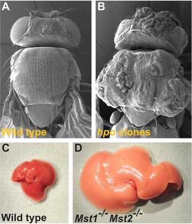
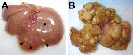
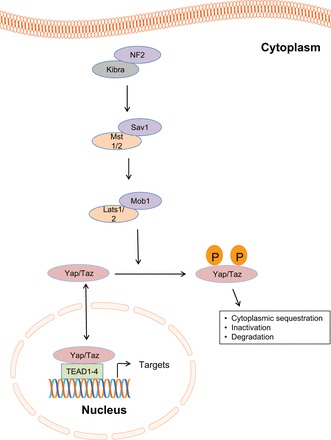
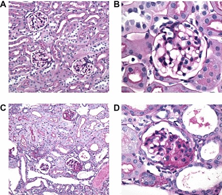
The Hippo signaling pathway is an evolutionarily conserved kinase cascade, playing multiple roles in embryonic development that controls organ size, cell proliferation, and apoptosis. At the center of this network lie the Hippo kinase target and downstream pathway effector Yes-associated protein (YAP) and its paralog TAZ. In its phosphorylated form, cytoplasmic YAP is sequestered in an inactive state. When it is dephosphorylated, YAP, a potent oncogene, is activated and relocates to the nucleus to interact with a number of transcription factors and signaling regulators that promote cell growth, differentiation, and survival. The identification of YAP activation in human cancers has made it an attractive target for chemotherapeutic drug development. Little is known to date about the function of the Hippo pathway in the kidney, but that is rapidly changing. Recent studies have shed light on the role of Hippo-YAP signaling in glomerular and lower urinary tract embryonic development, maintenance of podocyte homeostasis, the integrity of the glomerular filtration barrier, regulation of renal tubular cyst growth, renal epithelial injury in diabetes, and renal fibrogenesis. This review summarizes the current knowledge of the Hippo-YAP signaling axis in the kidney under normal and disease conditions.
Keywords: Hippo; kidney; podocyte.
Copyright © 2016 the American Physiological Society.
Figures
References
-
- Baumgartner R, Poernbacher I, Buser N, Hafen E, Stocker H. The WW domain protein Kibra acts upstream of Hippo in Drosophila. Dev Cell 18: 309–316, 2010. - PubMed
-
- Brancati FL, Whelton PK, Randall BL, Neaton JD, Stamler J, Klag MJ. Risk of end-stage renal disease in diabetes mellitus: a prospective cohort study of men screened for MRFIT. Multiple Risk Factor Intervention Trial. JAMA 278: 2069–2074, 1997. - PubMed
-
- Camargo FD, Gokhale S, Johnnidis JB, Fu D, Bell GW, Jaenisch R, Brummelkamp TR. YAP1 increases organ size and expands undifferentiated progenitor cells. Curr Biol 17: 2054–2060, 2007. - PubMed
Publication types
MeSH terms
Substances
Grants and funding
LinkOut - more resources
Full Text Sources
Other Literature Sources
Research Materials