. 2016 Jul 11;90(15):6784-98.
doi: 10.1128/JVI.00095-16. Print 2016 Aug 1.

Uukuniemi Virus as a Tick-Borne Virus Model

Affiliations

Uukuniemi Virus as a Tick-Borne Virus Model

Magalie Mazelier et al. J Virol. .

Abstract

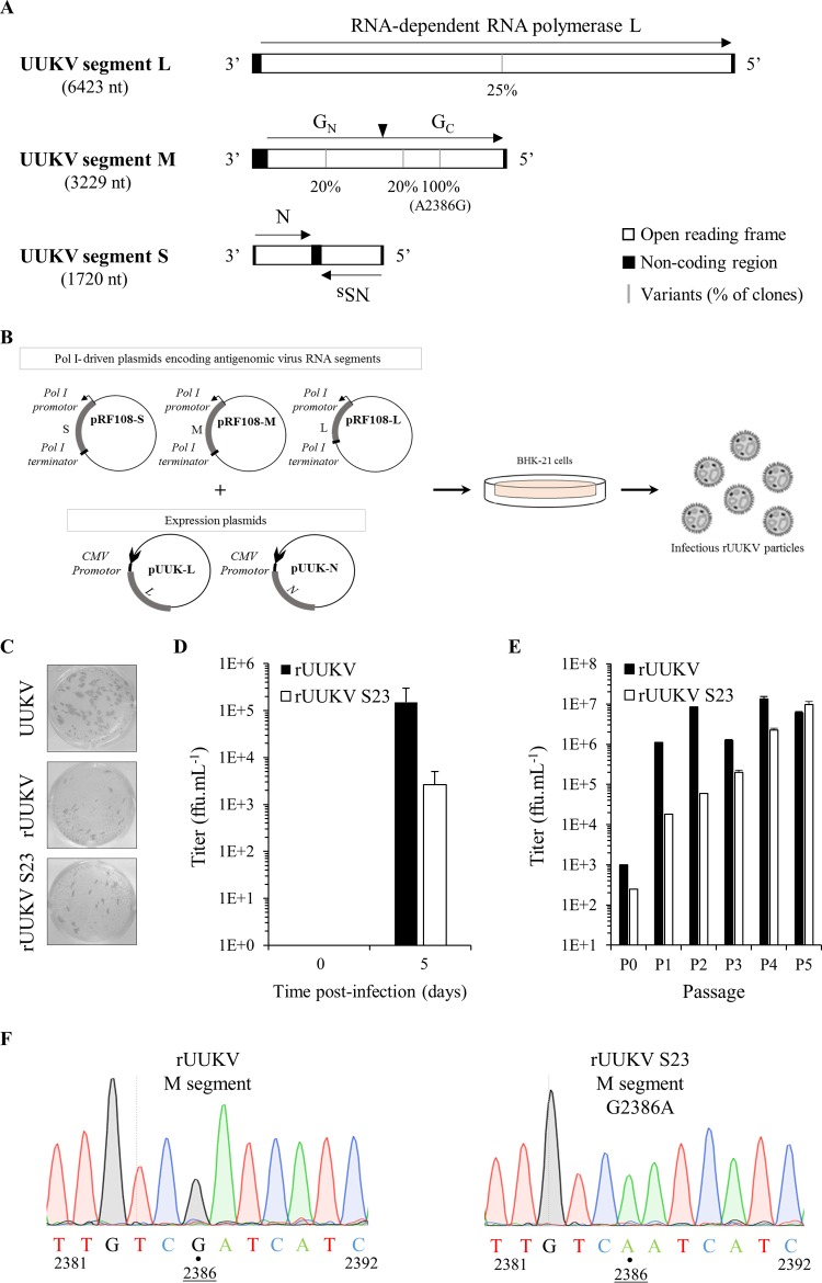
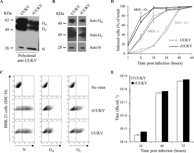
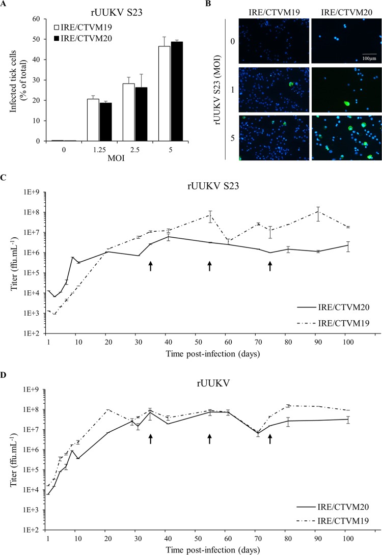
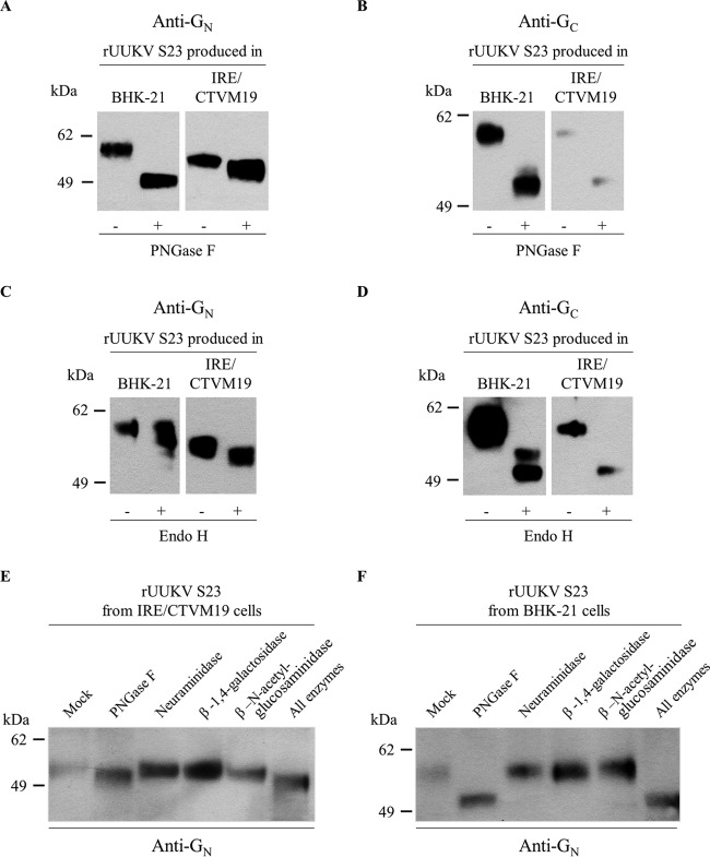
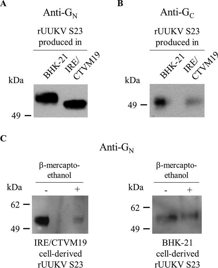
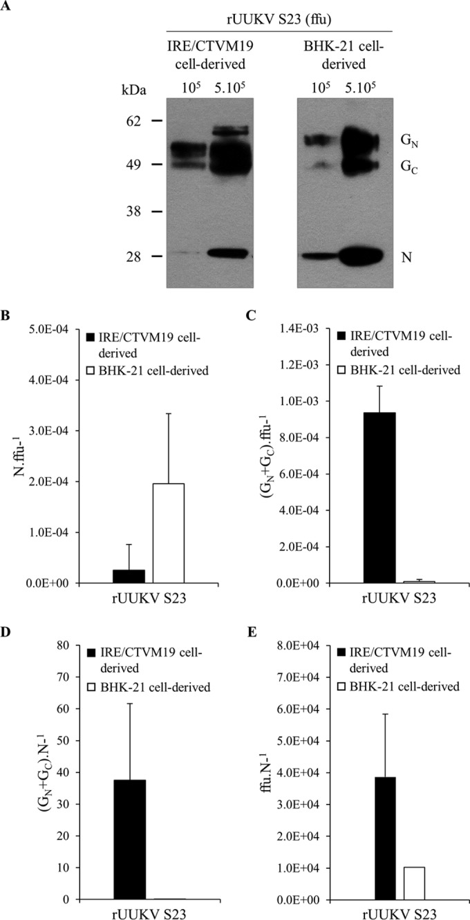
In the last decade, novel tick-borne pathogenic phleboviruses in the family Bunyaviridae, all closely related to Uukuniemi virus (UUKV), have emerged on different continents. To reproduce the tick-mammal switch in vitro, we first established a reverse genetics system to rescue UUKV with a genome close to that of the authentic virus isolated from the Ixodes ricinus tick reservoir. The IRE/CTVM19 and IRE/CTVM20 cell lines, both derived from I. ricinus, were susceptible to the virus rescued from plasmid DNAs and supported production of the virus over many weeks, indicating that infection was persistent. The glycoprotein GC was mainly highly mannosylated on tick cell-derived viral progeny. The second envelope viral protein, GN, carried mostly N-glycans not recognized by the classical glycosidases peptide-N-glycosidase F (PNGase F) and endoglycosidase H (Endo H). Treatment with β-mercaptoethanol did not impact the apparent molecular weight of GN On viruses originating from mammalian BHK-21 cells, GN glycosylations were exclusively sensitive to PNGase F, and the electrophoretic mobility of the protein was substantially slower after the reduction of disulfide bonds. Furthermore, the amount of viral nucleoprotein per focus forming unit differed markedly whether viruses were produced in tick or BHK-21 cells, suggesting a higher infectivity for tick cell-derived viruses. Together, our results indicate that UUKV particles derived from vector tick cells have glycosylation and structural specificities that may influence the initial infection in mammalian hosts. This study also highlights the importance of working with viruses originating from arthropod vector cells in investigations of the cell biology of arbovirus transmission and entry into mammalian hosts.

Importance: Tick-borne phleboviruses represent a growing threat to humans globally. Although ticks are important vectors of infectious emerging diseases, previous studies have mainly involved virus stocks produced in mammalian cells. This limitation tends to minimize the importance of host alternation in virus transmission to humans and initial infection at the molecular level. With this study, we have developed an in vitro tick cell-based model that allows production of the tick-borne Uukuniemi virus to high titers. Using this system, we found that virions derived from tick cells have specific structural properties and N-glycans that may enhance virus infectivity for mammalian cells. By shedding light on molecular aspects of tick-derived viral particles, our data illustrate the importance of considering the host switch in studying early virus-mammalian receptor/cell interactions. The information gained here lays the basis for future research on not only tick-borne phleboviruses but also all viruses and other pathogens transmitted by ticks.


Figures

FIG 1
FIG 1
Recovery of UUKV from Pol I-driven plasmid DNAs. (A) The trisegmented, negative-sense RNA genome of UUKV. The black arrowhead shows the cleavage site in the polyprotein precursor of the glycoproteins GN and GC. The black bars indicate the nucleotides found to be mutated in the UUKV laboratory strain relative to the sequence of UUKV strain 23. The penetrance of each mutation is indicated underneath. Four to five clones were analyzed for each viral genome segment. (B) Schematic representation of the Pol I-driven UUKV rescue system. (C) Focus-forming assay used for the titration of UUKV strains. Examples are shown for the UUKV lab strain (UUKV) and the viruses rescued from plasmid DNAs after five passages in BHK-21 cells (rUUKV and rUUKV S23). After 3 days of incubation at 37°C, foci were immunostained with the rabbit polyclonal antibody U2 against the viral proteins N, GN, and GC. (D) rUUKV and rUUKV S23 production 5 days after transfection of plasmids expressing L, M, and S segments under the control of the Pol I promoter together with the UUKV L and N expression plasmids pUUK-L and pUUK-N in BHK-21 cells. (E) Titer of rUUKV and rUUKV S23 after rescue (passage 0, P0) and up to five passages (P1 to P5) in BHK-21 cells. FFU, focus-forming units. (F) Sequence analysis of the rUUKV S23 M segment compared to that of rUUKV carried out from vRNA purified extracts after five passages in BHK-21 cells.
FIG 2
FIG 2
Characterization of UUKV rescued from plasmids. The UUKV lab strain and rUUKV were analyzed by SDS-PAGE and Western blotting under reducing conditions (A) using the rabbit polyclonal antibody U2 against the three structural viral proteins N, GN, and GC or under nonreducing conditions (B) with the mouse monoclonal antibodies 8B11A3, 6G9E5, and 3D8B3 that recognize each of the structural proteins N, GN, and GC, respectively. (C) BHK-21 cells were exposed to the UUKV lab strain or rUUKV at an MOI of 0.1 for 24 h. After fixation and permeabilization, infected cells were immunostained for N, GN, and GC with the mouse monoclonal antibodies 8B11A3, 6G9E5, and 3D8B3, respectively, and analyzed by flow cytometry. SSC-H, side scatter, height. (D) Infection of BHK-21 cells by UUKV and rUUKV was monitored over 64 h using the flow cytometry-based assay used for the experiment shown in panel C. Infection is given as the percentage of N protein-positive cells. (E) Supernatants collected from cells infected at an MOI of 0.1 and at indicated times were assessed for the production of infectious viral progeny by focus-forming assay.
FIG 3
FIG 3
Infection of tick cells by rUUKV and rUUKV S23 is persistent. (A) Tick cell lines IRE/CTVM19 and IRE/CTVM20 were exposed to BHK-21 cell-derived rUUKV S23 at the indicated MOIs for 48 h. Infection was analyzed by flow cytometry after immunostaining against the nucleoprotein N. (B) IRE/CTVM19 and IRE/CTVM20 cells were exposed to various MOIs of rUUKV S23 derived from BHK-21 cells. The next day, infected cells were immunostained for the intracellular UUKV nucleoprotein N using the anti-N primary mouse monoclonal antibody 8B11A3 and an AF488-coupled anti-mouse secondary monoclonal antibody (green). Nuclei were stained with Hoechst (blue), and samples were analyzed by wide-field microscopy. (C and D) IRE/CTVM19 and IRE/CTVM20 cells were exposed to rUUKV S23 and rUUKV, as indicated. Supernatant (200 μl) of infected cells was harvested daily during the first 10 days and every 10 days thereafter. The production of infectious viral particles in the supernatant was determined by focus-forming assays. The cells were subcultured in fresh complete medium (1:1) after sampling of the parent cells on days 34, 54, and 74 (black arrows).
FIG 4
FIG 4
The C type lectin DC-SIGN enhances infection of human cells by tick cell-derived rUUKV S23. (A) BHK-21 cells were infected (at an MOI of 0.1) with rUUKV S23 derived from IRE/CTVM19 cells for 18 h and immunostained for N, GN, and GC proteins prior to flow cytometry analysis. (B) Parental (Raji) and DC-SIGN-expressing Raji cells (Raji DC-SIGN+) were infected with IRE/CTVM19 cell-derived rUUKV S23 and analyzed by flow cytometry 16 h after immunostaining against the viral nucleoprotein. (C) Parental (HeLa) and DC-SIGN-expressing HeLa cells (HeLa DC-SIGN+) were exposed to various MOIs of IRE/CTVM19 cell-derived rUUKV S23. The next day, infected cells were immunostained for the intracellular virus nucleoprotein N using the anti-N primary mouse monoclonal antibody 8B11A3 and an AF488-coupled anti-mouse secondary monoclonal antibody (green). Nuclei were stained with Hoechst (blue), and samples were analyzed by wide-field microscopy. (D) Raji DC-SIGN-expressing cells were exposed to IRE/CTVM19 cell-derived rUUKV S23 (MOI of ∼1) in the presence of inhibitors blocking DC-SIGN, namely, EDTA (5 mM) or the neutralizing mouse monoclonal antibody mAb1621 (25 μg · ml−1). Intracellular viral antigens were detected by immunostaining with an anti-UUKV rabbit polyclonal antibody, followed by incubation with AF647-conjugated secondary antibodies. Infection was analyzed by flow cytometry 18 h later and normalized to infection of DC-SIGN-expressing Raji cells in the absence of inhibitor (as a percentage of the control).
FIG 5
FIG 5
Glycosylation of the rUUKV S23 envelope glycoproteins GN and GC on virions produced from tick and mammalian cells. IRE/CTVM19 and BHK-21 cell-derived rUUKV S23 purified through a 25% sucrose cushion was reduced and denatured before digestion with PNGase F (A, B, E, and F), Endo H (C and D), or neuraminidase, β-1,4-galactosidase, and β-N-acetylglucosaminidase (E and F). Proteins were analyzed by SDS-PAGE and Western blotting using the rabbit polyclonal antibodies K1224 and K5 against linear epitopes in the viral glycoproteins GN (A, C, E, and F) and GC (B and D), respectively.
FIG 6
FIG 6
Electrophoretic mobility of the glycoproteins GN and GC on rUUKV S23 produced in tick and mammalian cells. (A and B) BHK-21 and IRE/CTVM19 cell-derived rUUKV S23 purified through a 25% sucrose cushion were analyzed by nonreducing SDS-PAGE and Western blotting, using the mouse monoclonal antibodies 6G9E5 and 3D8B3 against conformational epitopes in GN (A) and GC (B), respectively. (C) rUUKV S23 viruses produced in BHK-21 and IRE/CTVM19 cells were purified through a 25% sucrose cushion and analyzed by nonreducing (−) or reducing (+) SDS-PAGE and Western blotting with the rabbit polyclonal anti-GN antibody K1224.
FIG 7
FIG 7
The structural rUUKV S23 proteins GN, GC, and N in infectious particles derived from tick and mammalian cells. (A) The amounts of viral glycoproteins and N protein for 105 and 5 × 10⁵ focus forming units (FFU) of purified rUUKV S23, produced in either tick cells or BHK-21 cells, were analyzed by SDS-PAGE and Western blotting using the rabbit polyclonal anti-UUKV antibody U2, which recognized GN, GC, and N. (B to E) The amount of viral glycoproteins and protein N for identical amounts of purified infectious rUUKV S23 was determined by quantitative Western blotting (Odyssey Imaging Systems) with the anti-UUKV U2 and an anti-rabbit infrared fluorescence secondary antibody. The ratios of the amounts of N protein (B) or viral glycoproteins (C) per FFU and the ratios of the amounts of viral glycoproteins (D) or number of FFU (E) per relative unit of N protein are shown.
FIG 8
FIG 8
Low-pH dependence of rUUKV S23 for infection. (A and B) Raji and HeLa cells that stably express DC-SIGN were pretreated with NH4Cl, a weak base that neutralizes the endosomal pH, and then exposed to IRE/CTVM19 cell-derived rUUKV S23 (MOI of ∼5) in the continuous presence of the inhibitor. Infected cells were harvested 16 h later and immunostained for the UUKV nucleoprotein N. Infection was analyzed by flow cytometry (A) or wide-field microscopy counting at least 200 cells in more than three independent fields (B). Data were normalized to DC-SIGN-expressing cells infected in the absence of inhibitor (as a percentage of the control). (C and D) IRE/CTVM19 and IRE/CTVM20 cells were infected with BHK-21 cell-derived rUUKV S23 at an MOI of 5 for 36 h in the continuous presence of NH4Cl. Infection was analyzed by flow cytometry and normalized against data obtained in the absence of inhibitor (as a percentage of the control).

References

    1. Leger P, Lozach PY. 2015. Bunyaviruses: from transmission by arthropods to virus entry into the mammalian host first-target cells. Future Virol 10:859–881. doi:10.2217/fvl.15.52. - DOI
    1. McMullan LK, Folk SM, Kelly AJ, MacNeil A, Goldsmith CS, Metcalfe MG, Batten BC, Albarino CG, Zaki SR, Rollin PE, Nicholson WL, Nichol ST. 2012. A new phlebovirus associated with severe febrile illness in Missouri. N Engl J Med 367:834–841. doi:10.1056/NEJMoa1203378. - DOI - PubMed
    1. Yu XJ, Liang MF, Zhang SY, Liu Y, Li JD, Sun YL, Zhang L, Zhang QF, Popov VL, Li C, Qu J, Li Q, Zhang YP, Hai R, Wu W, Wang Q, Zhan FX, Wang XJ, Kan B, Wang SW, Wan KL, Jing HQ, Lu JX, Yin WW, Zhou H, Guan XH, Liu JF, Bi ZQ, Liu GH, Ren J, Wang H, Zhao Z, Song JD, He JR, Wan T, Zhang JS, Fu XP, Sun LN, Dong XP, Feng ZJ, Yang WZ, Hong T, Zhang Y, Walker DH, Wang Y, Li DX. 2011. Fever with thrombocytopenia associated with a novel bunyavirus in China. N Engl J Med 364:1523–1532. doi:10.1056/NEJMoa1010095. - DOI - PMC - PubMed
    1. Liu S, Chai C, Wang C, Amer S, Lv H, He H, Sun J, Lin J. 2014. Systematic review of severe fever with thrombocytopenia syndrome: virology, epidemiology, and clinical characteristics. Rev Med Virol 24:90–102. doi:10.1002/rmv.1776. - DOI - PMC - PubMed
    1. Muehlenbachs A, Fata CR, Lambert AJ, Paddock CD, Velez JO, Blau DM, Staples JE, Karlekar MB, Bhatnagar J, Nasci RS, Zaki SR. 2014. Heartland virus-associated death in Tennessee. Clin Infect Dis 59:845–850. doi:10.1093/cid/ciu434. - DOI - PMC - PubMed

Publication types

LinkOut - more resources