. 2016 Jul 11;90(15):6864-6883.
doi: 10.1128/JVI.00469-16. Print 2016 Aug 1.

Both cis and trans Activities of Foot-and-Mouth Disease Virus 3D Polymerase Are Essential for Viral RNA Replication

Affiliations

Both cis and trans Activities of Foot-and-Mouth Disease Virus 3D Polymerase Are Essential for Viral RNA Replication

Morgan R Herod et al. J Virol. .

Abstract

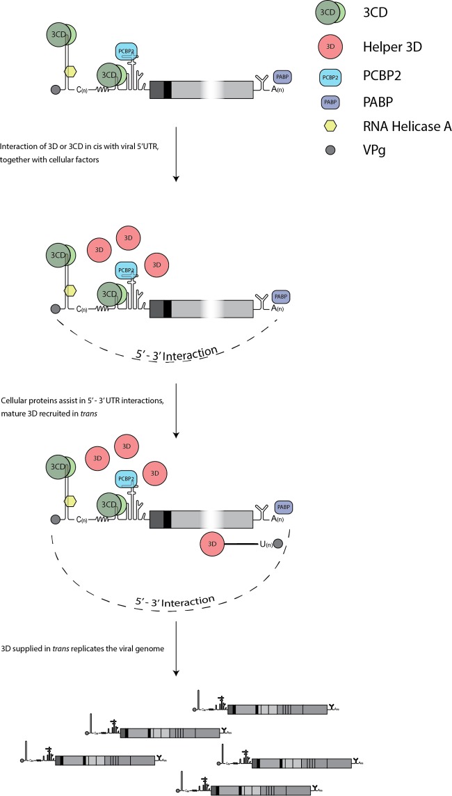
The Picornaviridae is a large family of positive-sense RNA viruses that contains numerous human and animal pathogens, including foot-and-mouth disease virus (FMDV). The picornavirus replication complex comprises a coordinated network of protein-protein and protein-RNA interactions involving multiple viral and host-cellular factors. Many of the proteins within the complex possess multiple roles in viral RNA replication, some of which can be provided in trans (i.e., via expression from a separate RNA molecule), while others are required in cis (i.e., expressed from the template RNA molecule). In vitro studies have suggested that multiple copies of the RNA-dependent RNA polymerase (RdRp) 3D are involved in the viral replication complex. However, it is not clear whether all these molecules are catalytically active or what other function(s) they provide. In this study, we aimed to distinguish between catalytically active 3D molecules and those that build a replication complex. We report a novel nonenzymatic cis-acting function of 3D that is essential for viral-genome replication. Using an FMDV replicon in complementation experiments, our data demonstrate that this cis-acting role of 3D is distinct from the catalytic activity, which is predominantly trans acting. Immunofluorescence studies suggest that both cis- and trans-acting 3D molecules localize to the same cellular compartment. However, our genetic and structural data suggest that 3D interacts in cis with RNA stem-loops that are essential for viral RNA replication. This study identifies a previously undescribed aspect of picornavirus replication complex structure-function and an important methodology for probing such interactions further.

Importance: Foot-and-mouth disease virus (FMDV) is an important animal pathogen responsible for foot-and-mouth disease. The disease is endemic in many parts of the world with outbreaks within livestock resulting in major economic losses. Propagation of the viral genome occurs within replication complexes, and understanding this process can facilitate the development of novel therapeutic strategies. Many of the nonstructural proteins involved in replication possess multiple functions in the viral life cycle, some of which can be supplied to the replication complex from a separate genome (i.e., in trans) while others must originate from the template (i.e., in cis). Here, we present an analysis of cis and trans activities of the RNA-dependent RNA polymerase 3D. We demonstrate a novel cis-acting role of 3D in replication. Our data suggest that this role is distinct from its enzymatic functions and requires interaction with the viral genome. Our data further the understanding of genome replication of this important pathogen.


Figures

FIG 1
FIG 1
Cartoon of the FMDV replicons used in complementation assays. Schematics of the FMDV subgenomic replicons used in this study. Complementation experiments involved simultaneous cotransfection of BHK-21 cells with equal amounts of replication-defective “mutant” RNA with helper replicon or yeast tRNA as a negative control. Replication of mutant and helper constructs was subsequently monitored by differential expression of fluorescent reporter transgenes encoding ptGFP and mCherry. Expression of ptGFP (A and B) and mCherry (C and D) was monitored hourly over a 24-h period. (A and C) Data are typically represented as mean positive cells per well ± standard deviations (SD) at 8 h posttransfection. (B and D) Time course of fluorescence expression over 20 h from a subset of samples (n = 3). The point mutation to the 3D active-site motif C (3D-GNN) has previously been shown to be recovered in trans by cotransfection with wild-type replicon.
FIG 2
FIG 2
Replication-defective 3D insertions can be recovered using wild-type helper replicon. (A) Cartoon of the FMDV replicon, including a schematic of the 3D sequence with conserved functional regions highlighted. The seven conserved motifs are colored as follows: A, red; B, green; C, yellow; D, light blue; E, orange; and F, dark blue. The region shown to interact with dsRNA is shaded in black. Complementation experiments involved cotransfecting mCherry replicons containing replication-defective 3D insertions, indicated with arrows, with wild-type ptGFP helper replicons or yeast tRNA. BHK-21 cells were seeded into 24-well plates and allowed to adhere for 16 h. Cells were cotransfected with mCherry replicons bearing replication-defective 3D insertions, a motif C active-site 3D mutation (3D-GNN), or a wild-type (wt) mCherry replicon control, together with wild-type ptGFP helper replicon or yeast tRNA as a negative control. Expression of mCherry (B) and ptGFP (C) was monitored hourly over a 24-h period. Data are represented as mean positive cells per well ± SD at 8 h posttransfection. Significance between plus ptGFP and plus yeast tRNA control (n = 3): *, P < 0.05; **, P < 0.01; ***, P < 0.001.
FIG 3
FIG 3
Replication-defective mutations in 3D motifs A and C can be recovered using wild-type helper replicon. (A) Cartoon of the FMDV replicon bearing replication-defective polymerase point mutations and a simplified version of the mCherry helper replicon (see Fig. 1 for full schematic). The schematic of the 3D polymerase gene is shown with the conserved motifs A to F highlighted. Arrows indicate the locations of mutated residues. Complementation assays involved cotransfection of ptGFP replicons bearing 3D point mutations with wild-type mCherry helper replicon or yeast tRNA. BHK-21 cells seeded into 24-well plates were cotransfected with ptGFP replicons harboring replication-defective 3D point mutations or a wild-type control, together with wild-type mCherry helper replicon or yeast tRNA as a negative control. Expression of ptGFP (B and D) and mCherry (C and E) were monitored hourly over a 24-h period. Data show mean positive cells per well ± SD (B and C) or mean total fluorescent intensity per well ± SD (D and E) at 8 h posttransfection. Significance between plus mCherry and plus yeast tRNA control (n = 2): *, P < 0.05; **, P < 0.01; ***, P < 0.001. (F) Diagrammatic representation of the 3D crystal structure showing the position of recoverable and nonrecoverable replication-defective point mutations as light green and hot pink spheres, respectively. Motif C containing the active-site GDD motif is colored yellow. Motifs A and F are colored red and dark blue, respectively.
FIG 4
FIG 4
Replication-defective 3D mutations do not affect RNA structure or 3D polyprotein processing. (A) Replicon RNAs containing codon-modified 3D were transfected into BHK-21 cells along with control replicons, and ptGFP expression was monitored hourly over 24 h. Data show mean numbers ± SD of ptGFP-positive cells per well at 8 h posttransfection (n = 2). (B) Effect of nocodazole on complementation. BHK-21 cells treated with 5 μM nocodazole or left untreated (see Materials and Methods for details) were cotransfected with wild-type or 3D-GNN ptGFP replicon RNA together with helper mCherry replicon or yeast tRNA negative control. ptGFP expression was monitored for 24 h. Data show normalized numbers of ptGFP-positive cells per well ± standard errors of the means (SEM) at 8 h posttransfection. Significance between results for untreated samples and samples treated with 5 μM nocodazole (n = 3) was determined: *, P < 0.05. (C) Western blot analysis of replicon DNA constructs transfected into BSR-T7 cells. Cells were seeded into 12-well plates, allowed to adhere for 16 h, and transfected with ptGFP replicon DNA constructs as indicated, and protein was harvested at 12 h posttransfection. Lysates were analyzed by Western blotting for 3D and GAPDH (glyceraldehyde-3-phosphate dehydrogenase) expression.
FIG 5
FIG 5
Wild-type helper 3D colocalizes with replication-defective mutant 3D. (A) Replicon RNAs containing C-terminal epitope labels were transfected into BHK-21 cells, and ptGFP expression was monitored hourly over a 24-h period. Data show mean numbers ± SD of ptGFP-positive cells per well at 8 h posttransfection (n = 2). (B) BHK-21 cells were cotransfected with FLAG-tagged mutant 3D replicons and HA-tagged 3D helper replicons, fixed at 4 h posttransfection, and labeled for FLAG-Alexa Fluor 568 (pseudocolored green) and HA-Alexa Fluor 647 (pseudocolored red) as described in Materials and Methods. Cell nuclei were counterstained with DAPI (blue). Images were captured on a Zeiss LSM-880 confocal microscope with Airyscan. Zoomed images represent the boxed areas on the merged images (bar, 20 μM). (C) Quantification of FLAG versus HA colocalization by confocal immunofluorescence. Fluorescent intensity of 3D-FLAG (green) versus 3D-HA (red) was plotted as two-dimensional (2D) scatterplots, and the number of costained pixels was determined by a 4-area gating set using controls. Data points show the percentages of coexpressing pixels from 8 randomly selected cells after cotransfection. Horizontal lines represents mean values ± SEM.
FIG 6
FIG 6
Replication-defective mutations to the 5′ UTR improve recovery of 3D mutations. (A) Schematic of the mutant ptGFP FMDV replicon genome and a simplified version of the mCherry genomes used in the reciprocal complementation assay. BHK-21 cells seeded into 24-well plates were cotransfected with replication-defective 3D mutant ptGFP replicons or wild-type control, together with mCherry replicons containing either an entire S-fragment deletion (ΔS) (B and D) or a replication-defective CRE mutation (CREA1G) (C and E). Wild-type mCherry helper replicon and yeast tRNA cotransfections were included as controls. Expression of ptGFP (B and C) and mCherry (D and E) was monitored hourly over a 24-h period. Data represent mean positive cells per well ± SD at 8 h posttransfection. Significance of differences between treatment and yeast tRNA negative control (n = 2): *, P < 0.05; **, P < 0.01; ***, P < 0.001.
FIG 7
FIG 7
Structure of the 3D-GC216/7AA mutant polymerase in complex with RNA. (A) Front view of a ribbon diagram of the 3D protein (gray) with the substituted amino acids A216 and A217 shown as light blue spheres. The seven conserved structure-sequence motifs are colored as follows: A, red; B, green; C, yellow; D, light blue; E, orange; F, blue; G, pink. (B) Electron density map around residues A216 and A217. Stereoview of a weighted 2Fo-Fc Fourier map, contoured at 1.5 σ, with the model placed inside (sticks colored in atom type code). (C) Superimposition of the wild-type (orange) and mutant (gray) 3D RNA structures. The regions showing the largest variations are highlighted as close-up insets: the mutated loop (left panel) and the entry of the template channel (right panel).
FIG 8
FIG 8
Expression of 3D alone is not sufficient for recovery of 3D mutations. (A) Western blot analysis of helper construct DNA transfected into BSR-T7 cells. Cells were seeded into 12-well plates, allowed to adhere for 16 h, and transfected with DNA constructs as indicated; protein was harvested at 12 h posttransfection. Lysates were analyzed by Western blotting for 3D and GAPDH expression. (B) For reciprocal complementation, BHK-21 cells seeded into 24-well plates were cotransfected with ptGFP replicons bearing the 3D-GNN replication-defective mutation or wild-type control, together with RNA transcripts expressing 3D alone or the entire 2A-3D polyprotein. Cotransfections were also performed with wild-type mCherry or mCherry-ΔS replicon transcripts and the equivalent 3D-GNN mutant constructs. Expression of ptGFP was monitored hourly over a 24-h period. Data represent mean positive cells per well ± SD at 8 h posttransfection. Significance compared to yeast tRNA control (n = 3): *, P < 0.05; **, P < 0.01; ***, P < 0.001.
FIG 9
FIG 9
Model of the cis and trans functions of 3D. 3D, either alone or as part of a larger precursor, interacts in cis with structural element within the 5′ UTR. In contrast, the polymerase functions of 3D can be readily supplied in trans. Cellular proteins are recruited at both the 5′ and 3′ UTRs. In addition, 5′-3′ RNA interactions are likely to be involved in the formation of an essential ribonucleoprotein complex.

Similar articles

Cited by

References

    1. Carrillo C, Tulman ER, Delhon G, Lu Z, Carreno A, Vagnozzi A, Kutish GF, Rock DL. 2005. Comparative genomics of foot-and-mouth disease virus. J Virol 79:6487–6504. doi:10.1128/JVI.79.10.6487-6504.2005. - DOI - PMC - PubMed
    1. Lopez de Quinto S, Martinez-Salas E. 1997. Conserved structural motifs located in distal loops of aphthovirus internal ribosome entry site domain 3 are required for internal initiation of translation. J Virol 71:4171–4175. - PMC - PubMed
    1. Lopez de Quinto S, Saiz M, de la Morena D, Sobrino F, Martinez-Salas E. 2002. IRES-driven translation is stimulated separately by the FMDV 3′-NCR and poly(A) sequences. Nucleic Acids Res 30:4398–4405. doi:10.1093/nar/gkf569. - DOI - PMC - PubMed
    1. Belsham GJ, Brangwyn JK. 1990. A region of the 5′ noncoding region of foot-and-mouth disease virus RNA directs efficient internal initiation of protein synthesis within cells: involvement with the role of L protease in translational control. J Virol 64:5389–5395. - PMC - PubMed
    1. Mason PW, Bezborodova SV, Henry TM. 2002. Identification and characterization of a cis-acting replication element (cre) adjacent to the internal ribosome entry site of foot-and-mouth disease virus. J Virol 76:9686–9694. doi:10.1128/JVI.76.19.9686-9694.2002. - DOI - PMC - PubMed

Publication types

Substances