. 2016 Jun 15;196(12):5036-46.
doi: 10.4049/jimmunol.1501973. Epub 2016 May 18.

Regulatory T Cell Dysfunction Acquiesces to BTLA+ Regulatory B Cells Subsequent to Oral Intervention in Experimental Autoimmune Encephalomyelitis

Affiliations

Regulatory T Cell Dysfunction Acquiesces to BTLA+ Regulatory B Cells Subsequent to Oral Intervention in Experimental Autoimmune Encephalomyelitis

Eduardo Huarte et al. J Immunol. .

Abstract

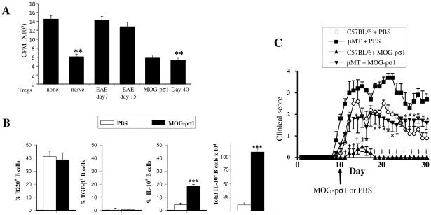
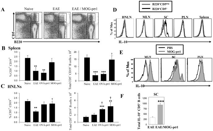
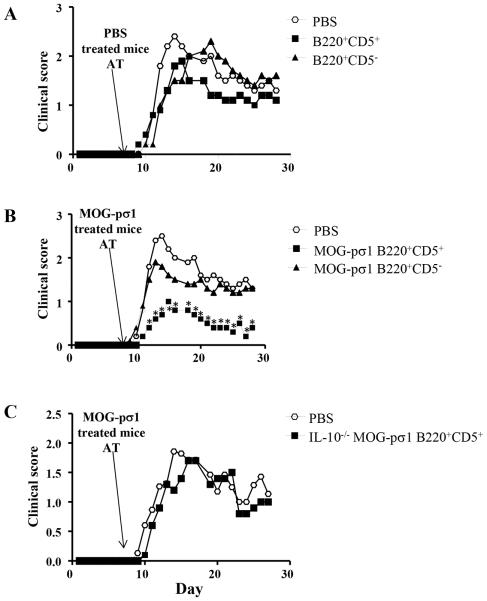
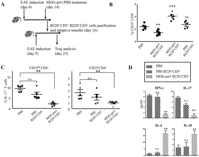
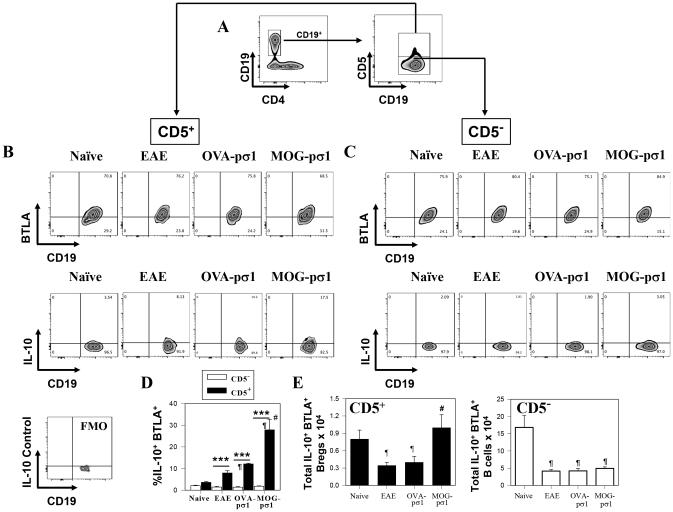
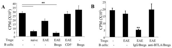
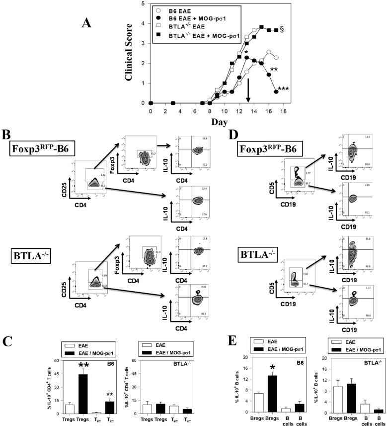
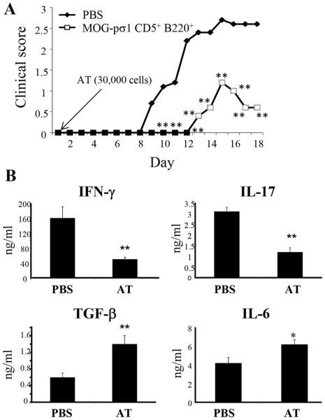
Regulatory T cells (Tregs) induced during autoimmunity often become quiescent and unable to resolve disease, suggesting inadequate activation. Resolution of established experimental autoimmune encephalomyelitis (EAE) can be achieved with myelin oligodendrocyte glycoprotein (MOG) fused to reovirus protein σ1 (MOG-pσ1), which activates Tregs, restoring protection, but requiring other regulatory cells to revitalize them. B cells have a dichotomous role in both the pathogenesis and recovery from EAE. Although inflammatory B cells contribute to EAE's pathogenesis, treatment of EAE mice with MOG-pσ1, but not OVA-pσ1, resulted in an influx of IL-10-producing B220(+)CD5(+) B regulatory cells (Bregs) enabling Tregs to recover their inhibitory activity, and in turn, leading to the rapid amelioration of EAE. These findings implicate direct interactions between Bregs and Tregs to facilitate this recovery. Adoptive transfer of B220(+)CD5(-) B cells from MOG-pσ1-treated EAE or Bregs from PBS-treated EAE mice did not resolve disease, whereas the adoptive transfer of MOG-pσ1-induced B220(+)CD5(+) Bregs greatly ameliorated EAE. MOG-pσ1-, but not OVA-pσ1-induced IL-10-producing Bregs, expressed elevated levels of B and T lymphocyte attenuator (BTLA) relative to CD5(-) B cells, as opposed to Tregs or effector T (Teff) cells, whose BTLA expression was not affected. These induced Bregs restored EAE Treg function in a BTLA-dependent manner. BTLA(-/-) mice showed more pronounced EAE with fewer Tregs, but upon adoptive transfer of MOG-pσ1-induced BTLA(+) Bregs, BTLA(-/-) mice were protected against EAE. Hence, this evidence shows the importance of BTLA in activating Tregs to facilitate recovery from EAE.


Figures

Figure 1
Figure 1
EAE derived Tregs are dysfunctional and B cells modulate EAE. (A) Tregs were purified from naïve or EAE mice (day 7 or 15 p.ch., scores 1 and 3 respectively), MOG-pσ1-treated mice (day 15 p.ch.), those naturally recovering from EAE (day 40, scores 0-1), and CD25 CD4+ Teff cells from naïve mice. Tregs were then co-cultured with Teff cells (Tregs/effector ratio1:2) in the presence of anti-CD3 plus anti-CD28 mAbs for 4 days before measuring extent of proliferation using a 3H-thymidine incorporation assay. A representative experiment of 5 is shown; **p ≤ 0.01 versus naïve cell proliferation. (B) C57BL/6 mice (8/group) were induced with MOG35-55 EAE, and 10 days later, treated with PBS or MOG-pσ1. The percentage and total combined LN and splenic B220+ B cells expressing TGF-β+ or IL-10 from individual mice was measured 20 days p.ch.; ***p < 0.001 versus PBS-treated EAE mice. (C) C57BL/6 or B cell-deficient μMT mice were orally dosed with 50 μg MOG-pσ1 or PBS 10 days after EAE challenge and monitored daily for disease course. An average of 5 mice per group is represented; *p < 0.05 versus PBS-dosed μMT, p < 0.05 versus PBS-dosed C57BL/6 mice.
Figure 2
Figure 2
MOG-pσ1 intervention promotes the development of regulatory B cells (Bregs). EAE was induced on C57BL/6 mice, and 14 days later, mice were treated with PBS or MOG-pσ1. (A) A representative dot-plot showing the percentage of B220+CD5+ Bregs is depicted. Bar graphs indicate mean (± SEM) percentages of and total (B) splenic and (C) HNLN CD19+CD5+ B cells (9 mice/group) in naïve mice, EAE mice, and EAE mice treated with OVA-pσ1 or MOG-pσ1; ***p < 0.001, **p ≤ 0.01, *p < 0.05 versus naïve B cells. (D) EAE mice were evaluated d14 p.ch. for IL-10 production by B220+CD5+ and B220+CD5neg cells in HNLNs, MLNs, SCs, PLNs, and spleens. (E) EAE (day 14 p.ch.) mice (5/group) were analyzed 24h after PBS or MOG-pσ1 treatment for intracellular IL-10 production for pooled SCs, MLNs, and PLNs. (F) The total number of IL-10-producing CD5+ B cells from the SCs of PBS- and MOG-pσ1-treated EAE mice is depicted (right panel); ***p ≤0.001 vs. EAE mice. A representative experiment of three is shown.
Figure 3
Figure 3
MOG-pσ1-, not PBS-induced Bregs, confers protection against EAE. C57BL/6 or IL-10−/− mice (10/group) were induced with EAE and 2 weeks later treated with PBS or MOG-pσ1. Twenty four hours after MOG-pσ1 treatment, combined LNs and splenic B cells were sorted into B220+CD5 B cells and B220+CD5+ B cell subsets, and 2.0x10⁶ B cells were adoptively transferred (AT) into C57BL/6 recipients (5/group) that were already induced with EAE 7 days earlier. (A) PBS-induced B220+CD5 B cells or Bregs failed to ameliorate EAE. (B) MOG-pσ1-induced Bregs, not B220+CD5 B cells were able to significantly reduce EAE severity. (C) In the same experiment, IL-10−/− B220+CD5+ Bregs induced with MOG-pσ1 failed to significantly reduce EAE, indicating that their effect is IL-10-mediated. Panels A and B share the same control PBS group. *p < 0.01 versus PBS-treated mice or recipients adoptively transferred with MOG-pσ1-induced B220+ CD5 B cells.
Figure 4
Figure 4
Adoptive transfer of MOG-pσ1-treated B220+CD5+ cells increases both the frequency and activity of CD25+CD4+Tregs in recipient mice. C57BL/6 mice (10/group) were treated with MOG-pσ1 or PBS 2 wks after EAE induction, and B220+CD5+ and B220+CD5 cells were purified 48 h after treatment from combined LNs and spleens. B cells (105 cells/mouse) were adoptively transferred into C57BL/6 mice (5/group) previously induced with EAE 7 days earlier. (A) A schematic depicting the experiment is provided. (B) The percentage of CD25+CD4+ T cells in each group was measured 1 wk later. Only recipients given MOG-pσ1-induced B220+CD5+ Bregs showed an increase in the percentage of CD25+CD4+ T cells; ***p = 0.002, **p < 0.01 versus PBS-treated mice; ns, not significant. (C) Analysis of the percentage of IL-17-expressing cells between CD25HiCD4+ and CD25 CD4+ T cells. Recipients given MOG-pσ1-induced Bregs showed significantly fewer IL-17-producing cells; **p < 0.01; ns, not significant. (D) Bregs stimulate Tregs to produce IL-4 and IL-10 and suppress IFN-γ and IL-17 production. CD25+CD4+ T cells were purified from PBS-treated mice and recipients adoptively transferred with PBS- or MOG-pσ1-induced B220+CD5+ Bregs, and stimulated in vitro with anti-CD3 + anti-CD28 mAbs. Cytokine levels (mean ± SEM from triplicate cultures) in culture supernatants were measured by ELISA 4 days after culture, and values corrected for spontaneous production from unstimulated cells; **p < 0.01 versus PBS-treated mice; ns, not significant. Data are representative from two experiments.
Figure 5
Figure 5
MOG-pσ1 intervention upon EAE promotes increased BTLA expression/activation of Bregs for enhanced IL-10 production. EAE was induced in C57BL/6 mice, and mice were treated with PBS, OVA-pσ1, or MOG-pσ1 at the peak of disease (day 14), and splenic B cells were subsequently evaluated for BTLA expression. (A) Gating strategy used to identify Bregs staining for CD19 vs. CD4, and CD19+ B cells further analyzed for CD5 expression. (B) CD5+ and (C) CD5 CD19+ B cells from naïve, PBS-treated EAE, and MOG-pσ1-treated EAE mice were analyzed for BTLA and IL-10 expression; a fluorescence minus one (FMO) control histogram was performed for intracellular IL-10 staining. (D) The percent and (E) total IL-10+ BTLA+ CD5+ or CD5 B cells ± SEM from individual mice from naïve (n=9), PBS- (n=11), OVA-pσ1- (n=9), and MOG-pσ1-treated (n=11) mice are shown; ***p < 0.001, *p < 0.05 versus the corresponding CD5 B cell subset; #p < 0.005 versus EAE mice; and p < 0.05 versus naïve mice.
Figure 6
Figure 6
Bregs activate EAE Tregs to restore Treg function. (A, B) To test if Bregs can reactivate Tregs in vitro, (A) splenic Tregs obtained from naïve or EAE mice were stimulated as described in Fig. 1A, co-cultured with naïve CD4+ T cells in the absence or presence of CD19+ CD5+ Bregs or CD19+ CD5 B cells obtained from MOG-pσ1-treated EAE mice, and measured their 3H-thymidine incorporation 4 days later (Tregs/B cells/Teff cells ratio1:1:2). As a control, naïve CD4+ T cell proliferation in the presence of B220+ CD5+ cells (without Tregs) was also included. An average of 6 replicates/treatment group is depicted, and these data are representative of three experiments; **p ≤ 0.01 versus naïve T cell proliferation. (B) Tregs obtained from naïve or EAE mice were co-cultured with naïve CD4+ T cells in the presence or absence of B220+CD5+ Bregs (Tregs/Bregs/Teff cells ratio1:1:2), and the extent of CD4 T cell proliferation was measured 4 days later. Where indicated, B220+CD5+ Bregs were pretreated with an anti-BTLA mAb. An average of 6 replicates/treatment group is depicted, and these data are representative of two experiments; **p ≤ 0.01 versus naïve T cell proliferation.
Figure 7
Figure 7
A lack of IL-10 activation in BTLA−/− accounts for increased susceptibility to MOG-induced EAE. (A) Groups of C57BL/6 and BTLA−/− mice were induced with EAE on day 0, and were subsequently treated with PBS (B6 mice, n=7; BTLA−/− mice, n = 6) or MOG-pσ1 (B6 mice, n=7; BTLA−/− mice, n = 6) on day 13 (arrow). Clinical scores were measured throughout the disease; ***p < 0.001, **p < 0.005, *p = 0.036 versus PBS-treated B6 mice; §p < 0.030 for PBS- or MOG-pσ1-treated BTLA−/− mice versus PBS-treated B6 mice. (B) MOG-pσ1-treated BTLA−/− mice failed to show an induction of IL-10. (B-E) Additional groups of PBS- and MOG-pσ1-treated Foxp3RFP-B6 and BTLA−/− mice were treated as described in (A), and four days after treatment, isolated HNLNs were evaluated for IL-10 expression by (B and C) Tregs and Teff cells and (D and E) CD5+ Bregs and CD5 B cells. In BTLA-sufficient mice, MOG-pσ1 was able to significantly induce IL-10 by both (C) Tregs and (E) Bregs while no changes in IL-10 production by these same cells from BTLA−/− mice remained unchanged; **p = 0.003, *p ≤ 0.019 versus cells from EAE mice. Depicted data are representative of two experiments.
Figure 8
Figure 8
Adoptive transfer of Bregs restores protection to EAE in BTLA−/− mice. (A) BTLA+ CD19+CD5+ Bregs were purified from MOG-pσ1-treated C57BL/6 mice and 3x10⁴ Bregs were adoptively transferred (AT) into BTLA−/− mice at the time of EAE induction; clinical scores were measured throughout the disease (5/group, **p ≤ 0.01). (B) Cytokine analyses were performed on purified splenic CD4+ T cells at the peak of the disease (day 18 p.ch.). Purified CD4+ T cells were cultured for 4 days in the presence of MOG35-55 (5 μg/ml) and irradiated APCs. Cytokine production showed significant elevations in TGF-β and IL-6 when compared to PBS mice with concomitant reductions in IFN-γ and IL-17 production (* p < 0.05, **p ≤ 0.01).

Similar articles

Cited by

References

    1. Edwards JC, G. Cambridge Sustained improvement in rheumatoid arthritis following a protocol designed to deplete B lymphocytes. Rheumatology (Oxford) 2001;40:205–211. - PubMed
    1. Looney RJ, Anolik JH, Campbell D, Felgar RE, Young F, Arend LJ, Sloand JA, Rosenblatt J, Sanz I. B cell depletion as a novel treatment for systemic lupus erythematosus: a phase I/II dose-escalation trial of rituximab. Arthritis Rheum. 2004;50:2580–2589. - PubMed
    1. Hauser SL, Waubant E, Arnold DL, Vollmer T, Antel J, Fox RJ, Bar-Or A, Panzara M, Sarkar N, Agarwal S, Langer-Gould A, Smith CH, H. T. Group B-cell depletion with rituximab in relapsing-remitting multiple sclerosis. N Engl J Med. 2008;358:676–688. - PubMed
    1. Evans JG, Chavez-Rueda KA, Eddaoudi A, Meyer-Bahlburg A, Rawlings DJ, Ehrenstein MR, Mauri C. Novel suppressive function of transitional 2 B cells in experimental arthritis. J Immunol. 2007;178:7868–7878. - PubMed
    1. Kala M, Rhodes SN, Piao WH, Shi FD, Campagnolo DI, Vollmer TL. B cells from glatiramer acetate-treated mice suppress experimental autoimmune encephalomyelitis. Exp Neurol. 2010;221:136–145. - PubMed

Publication types

MeSH terms