. 2016 May 18;23(6):278-88.
doi: 10.1101/lm.041822.116. Print 2016 Jun.

Retrosplenial cortex is required for the retrieval of remote memory for auditory cues

Affiliations

Retrosplenial cortex is required for the retrieval of remote memory for auditory cues

Travis P Todd et al. Learn Mem. .

Abstract

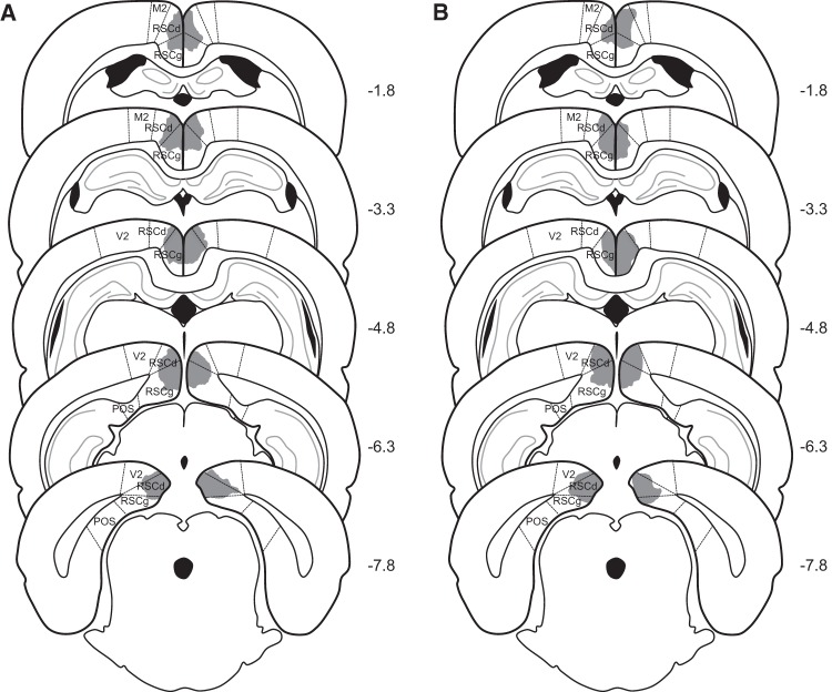
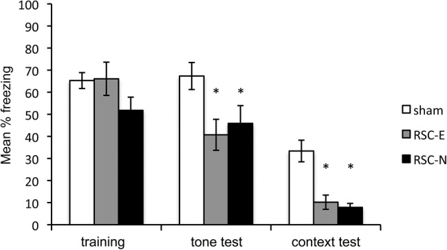
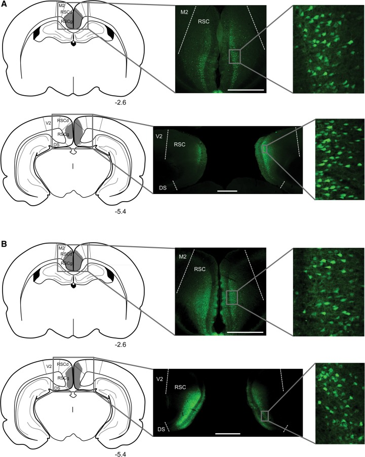
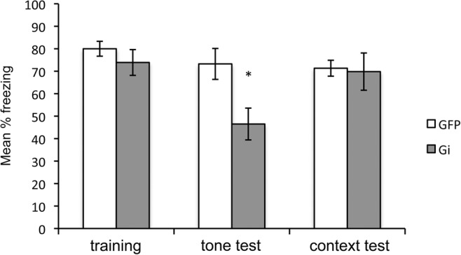
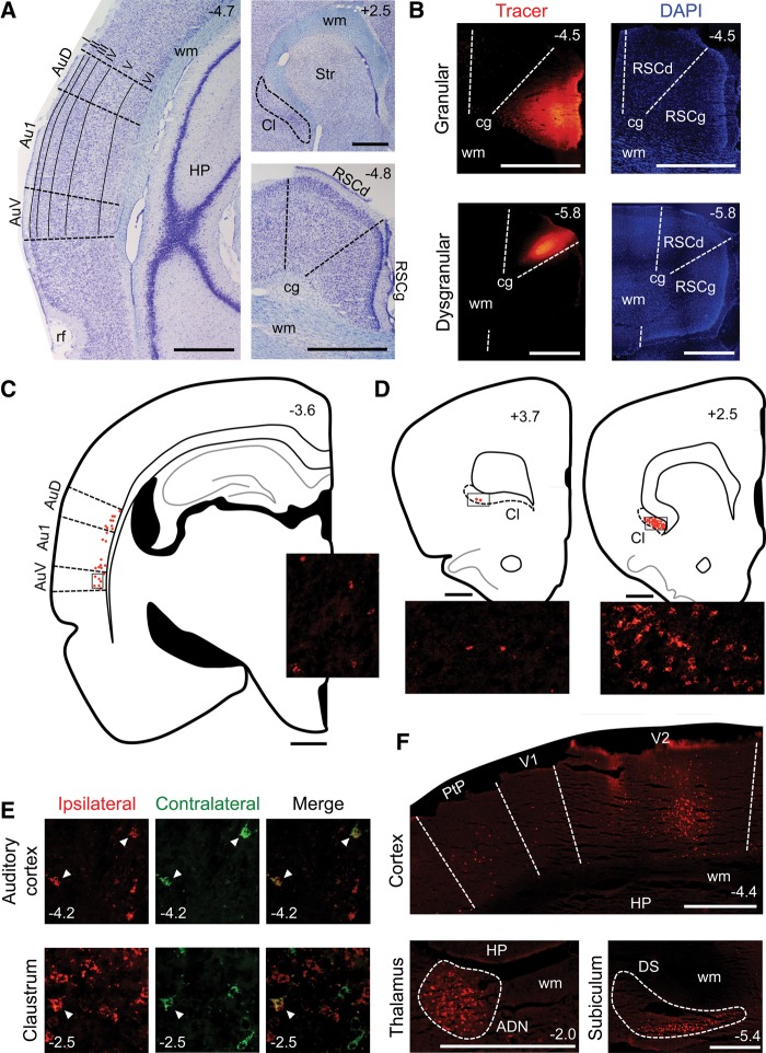
The restrosplenial cortex (RSC) has a well-established role in contextual and spatial learning and memory, consistent with its known connectivity with visuo-spatial association areas. In contrast, RSC appears to have little involvement with delay fear conditioning to an auditory cue. However, all previous studies have examined the contribution of the RSC to recently acquired auditory fear memories. Since neocortical regions have been implicated in the permanent storage of remote memories, we examined the contribution of the RSC to remotely acquired auditory fear memories. In Experiment 1, retrieval of a remotely acquired auditory fear memory was impaired when permanent lesions (either electrolytic or neurotoxic) were made several weeks after initial conditioning. In Experiment 2, using a chemogenetic approach, we observed impairments in the retrieval of remote memory for an auditory cue when the RSC was temporarily inactivated during testing. In Experiment 3, after injection of a retrograde tracer into the RSC, we observed labeled cells in primary and secondary auditory cortices, as well as the claustrum, indicating that the RSC receives direct projections from auditory regions. Overall our results indicate the RSC has a critical role in the retrieval of remotely acquired auditory fear memories, and we suggest this is related to the quality of the memory, with less precise memories being RSC dependent.


Figures

Figure 1.
Figure 1.
Schematic diagrams depicting the extent of representative electrolytic (A) and neurotoxic (B) lesions of the RSC in Experiment 1. The numbers adjacent to each section indicate the A/P position in mm relative to bregma based on Paxinos and Watson (2009). (M2) secondary motor cortex; (RSCd) retrosplenial cortex, dysgranular; (RSCg) retrosplenial cortex, granular; (V2) secondary visual cortex; (POS) post-subiculum.
Figure 2.
Figure 2.
Results of Experiment 1. Freezing behavior during the prelesion training session and the post-lesion remote tone and remote context test sessions. Training = mean percentage freezing during three post-shock periods. Tone test = mean percentage freezing during 20 shock-free presentations of the tone in Context B. Context test = mean percentage freezing during the 10-min test in Context A. (*) P < 0.05.
Figure 3.
Figure 3.
Virus expression in anterior and posterior portions of the RSC in a rat from group Gi (A) and a rat from group GFP (B) in Experiment 2. Schematic diagrams in the left column depict the extent of virus expression within the RSC, with the numbers below each section indicating the A/P position in mm relative to bregma based on Paxinos and Watson (2009). Low magnification (middle column) and high magnification (right column) images show virus-expressing cells. All scale bars represent 1 mm. (M2) secondary motor cortex; (RSCd) retrosplenial cortex, dysgranular; (RSCg) retrosplenial cortex, granular; (V2) secondary visual cortex; (DS) dorsal subiculum.
Figure 4.
Figure 4.
Results of Experiment 2. Freezing behavior during the training session and remote tone and remote context tests. Training = mean percentage freezing during three post-shock periods. Tone test = mean percentage freezing during 20 shock-free presentations of the tone in Context B. Context test = mean percentage freezing during the 10 min test in Context A. Prior to the remote tone and context tests, all rats were injected with CNO. Gi = AAV-hSyn-HA-hM4Di-IRES-mCitrine and GFP = AAV-hSyn-GFP. (*) P < 0.05
Figure 5.
Figure 5.
Results of Experiment 3. (A) Nissl-stained sections containing the auditory cortex (left), claustrum (upper right), or RSC (lower right). (B) Sections containing a tracer injection in the granular (top) or dysgranular (bottom) region of the RSC. The left column shows the tracer and the right column shows the DAPI stain. (C) Tracing of a section illustrating labeling in the auditory cortex following a tracer injection in the ipsilateral RSC. Each red dot represents one labeled cell. Inset image shows labeled cells in the region of the auditory cortex indicated by the rectangular outline on the tracing. (D) Tracings of sections illustrating labeling in anterior (left) and posterior (right) portions of the claustrum following a tracer injection in the ipsilateral RSC. Each red dot represents one labeled cell. Inset images show labeled cells in the regions of the claustrum indicated by the rectangular outlines on the tracings. (E) Bilaterally projecting cells in the auditory cortex (top) and claustrum (bottom). The left column shows cells projecting to the ipsilateral hemisphere and the middle column shows cells projecting to the contralateral hemisphere; these two images are merged in the right column. White arrows indicate double-labeled (i.e., bilaterally projecting) cells. (F) Other cortical and subcortical labeling patterns. The top image shows labeled cells in the primary and secondary visual cortices and the posterior parietal cortex, the lower left image shows labeled cells in the anterodorsal thalamic nucleus (outlined by the dashed line) and the lower right image shows labeled cells in the dorsal subiculum (outlined by the dashed line). The numbers in the corner of each image indicate the A/P position in mm relative to bregma, based on Paxinos and Watson (2009). All scale bars represent 1 mm. (Au1) primary auditory cortex; (AuD) secondary auditory cortex, dorsal; (AuV) secondary auditory cortex, ventral; (rf) rhinal fissure; (wm) white matter; (HP) hippocampus; (Cl) claustrum; (Str) striatum; (RSCg) retrosplenial cortex, granular; (RSCd) retrosplenial cortex, dysgranular; (cg) cingulum; (V1) primary visual cortex; (V2) secondary visual cortex; (PtP) parietal cortex, posterior; (ADN) anterodorsal thalamic nucleus; (DS) dorsal subiculum.

References

    1. Anagnostaras SG, Maren S, Fanselow MS. 1999. Temporally graded retrograde amnesia of contextual fear after hippocampal damage in rats: within-subject examination. J Neurosci 19: 1106–1114. - PMC - PubMed
    1. Arenos JD, Musty RE, Bucci DJ. 2006. Blockade of cannabinoid CB1 receptors alters contextual learning and memory. Eur J Pharmacol 539: 177–183. - PubMed
    1. Armbruster BN, Li X, Pausch MH, Hertzile S, Roth BL. 2007. Evolving the lock to fit the key to create a family of G protein-coupled receptors potently activated by an inert ligand. Proc Natl Acad Sci 104: 5163–5168. - PMC - PubMed
    1. Beneyto M, Prieto JJ. 2001. Connections of the auditory cortex with the claustrum and the endopiriform nucleus in the cat. Brain Res Bull 54: 485–498. - PubMed
    1. Blanchard RJ, Blanchard DC. 1969. Crouching as an index of fear. J Comp Physiol Psychol 67: 370–375. - PubMed

LinkOut - more resources