Review
. 2016 Aug;100(2):291-304.
doi: 10.1189/jlb.5RU0216-056R. Epub 2016 May 18.

Big insights from small volumes: deciphering complex leukocyte behaviors using microfluidics

Affiliations
Review

Big insights from small volumes: deciphering complex leukocyte behaviors using microfluidics

Daniel Irimia et al. J Leukoc Biol. 2016 Aug.

Abstract

Inflammation is an indispensable component of the immune response, and leukocytes provide the first line of defense against infection. Although the major stereotypic leukocyte behaviors in response to infection are well known, the complexities and idiosyncrasies of these phenotypes in conditions of disease are still emerging. Novel tools are indispensable for gaining insights into leukocyte behavior, and in the past decade, microfluidic technologies have emerged as an exciting development in the field. Microfluidic devices are readily customizable, provide tight control of experimental conditions, enable high precision of ex vivo measurements of individual as well as integrated leukocyte functions, and have facilitated the discovery of novel leukocyte phenotypes. Here, we review some of the most interesting insights resulting from the application of microfluidic approaches to the study of the inflammatory response. The aim is to encourage leukocyte biologists to integrate these new tools into increasingly more sophisticated experimental designs for probing complex leukocyte functions.

Keywords: dendritic cells; inflammation; monocytes; nanotechnology; neutrophils.


Figures

Figure 1.
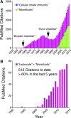
Figure 1.. Microfluidics and innate immunity, two fields of exponential growth.
(A) Graph shows PubMed citations/yr in response to the search terms “Cellular innate immunity” (magenta columns) and “Microfluidic” (green columns). Invention of the Boyden chamber corresponds to a surge in research in the field, peaking in the mid-1980s. Since its resurgence in the early 2000s, the field of leukocyte biology continues to expand exponentially. Following the first microfluidic prototypes for biomedical applications in the early 2000s, this area of study also exhibits exponential growth. (B) Graph shows PubMed citations/yr in response to the search term “Leukocyte” in combination with “Microfluidic” (magenta columns). Use of microfluidic assays to measure leukocyte behavior is becoming increasingly common, with ∼60% of publications emerging in the last 5 yr.
Figure 2.
Figure 2.. Models of focal leukocyte chemotaxis.
(A) Spinning disk confocal intravital microscopy of mouse neutrophils (lys-EGFP green) responding to a localized thermal injury (propidium iodide; red) on the surface of the liver from the Kubes lab. [Reproduced from McDonald, B., Pittman, K., Menezes, G. B., Hirota, S. A., Slaba, I., Waterhouse, C. C., Beck, P. L., Muruve, D. A., Kubes, P. (2010) Intravascular danger signals guide neutrophils to sites of sterile inflammation. Science 330, 362–366. Reprinted with permission from the American Association for the Advancement of Science (AAAS).] (B) A microfluidic model of focal chemotaxis allows parallel imaging of leukocyte chemotaxis from a central cell-loading chamber toward 16 identical chemokine reservoirs (red dye) on a single device. [Reproduced from Jones, C. N., Dalli, J., Dimisko, L., Wong, E., Serhan, C. N., Irimia, D. (2012) Microfluidic chambers for monitoring leukocyte trafficking and humanized nano-proresolving medicines interactions. Proc Natl Acad Sci USA 109, 20560–20565.] (C) Integration of a RBC filter facilitates use of whole-blood samples. Neutrophils (blue nuclei, Hoechst) are able to navigate the filter and migrate toward the chemokine source. [Reproduced from Hoang, A. N., Jones, C. N., Dimisko, L., Hamza, B., Martel, J., Kojic, N., Irimia, D. (2013) Measuring neutrophil speed and directionality during chemotaxis, directly from a droplet of whole blood. Technology 1, 49–57. Electronic version of an article published as Technology 1, 49–57 doi: 10.1142/S2339547813500040, copyright World Scientific Publishing Company, www.worldscientific.com.] (D) Coordinated recruitment of multiple cell types, such as neutrophils (blue) and macrophages (red) in response to complex signals, can be studied under defined conditions. [Reproduced from Jones, C. N., Dalli, J., Dimisko, L., Wong, E., Serhan, C. N., Irimia, D. (2012) Microfluidic chambers for monitoring leukocyte trafficking and humanized nano-proresolving medicines interactions. Proc Natl Acad Sci USA 109, 20560–20565.]
Figure 3.
Figure 3.. Modeling of inflammation resolution by neutrophil reverse migration.
(A) A transgenic zebrafish model of sterile inflammation. Neutrophils are labeled by GFP expression, driven by the myeloperoxidase promoter, allowing in vivo tracking of neutrophil migration relative to the wound (dashed yellow line) during recruitment and resolution phases. (B, i) Reverse-migrating neutrophils can be more easily visualized by spatially restricted photoconversion (from green to red) of wound-associated neutrophils (dashed white outline) that express the fluorescent protein Kaede. (ii) Reverse migration of neutrophils can then be monitored by identifying those cells that return to the tissue (white arrowhead). [A and B reproduced from Robertson, A. L., Holmes, G. R., Bojarczuk, A. N., Burgon, J., Loynes, C. A., Chimen, M., Sawtell, A. K., Hamza, B., Willson, J., Walmsley, S. R., Anderson, S. R. (2014) A zebrafish compound screen reveals modulation of neutrophil reverse migration as an anti-inflammatory mechanism. Sci Transl Med 6, 225ra29–225ra29. Reprinted with permission from AAAS.] (C) A microfluidic device to measure neutrophil recruitment and reverse migration from a drop of whole blood. (D, i) Neutrophils (blue tracks) are recruited (black arrowheads) from the whole blood to the chemotaxis chambers and then reverse migrate (blue arrowheads) down the chemokine gradient and back into the whole-blood reservoir. (ii) In the presence of microbial particles (zymosan; green), neutrophils are retained (red dots) at the chemokine reservoir and show reduced reverse migration. [C and D reproduced from Hamza, B., Irimia, D. (2015) Whole blood human neutrophil trafficking in a microfluidic model of infection and inflammation. Lab Chip 15, 2625–2633. Reprinted with permission from the Centre National de la Recherche Scientifique (CNRS) and The Royal Society of Chemistry.]
Figure 4.
Figure 4.. Modeling complex cell migration signatures.
(A, i) Two-photon intravital imaging of CD4 (GFP; green) and CD8 (DsRed; red) T cells in the epidermis (collagen visualized in blue using second-harmonic generation) following infection with HSV. (ii) 3D tracking of cell migration over time demonstrates the complexity of cell migration in vivo. [Reproduced from Gebhardt, T., Whitney, P. G., Zaid, A., Mackay, L. K., Brooks, A. G., Heath, W. R., Carbone, F. R., Mueller, S. N. (2011) Different patterns of peripheral migration by memory CD4+ and CD8+ T cells. Nature 477, 216–219, copyright 2011. Reprinted with permission from Macmillan Publishers Ltd., Nature 2011.] (B, i) A microfluidic device for studying complex cell migration signatures. Lymphocytes migrate through a maze of posts toward a chemokine reservoir. (ii) Cell migration (colored tracks) can be tracked at cellular resolution in 2 dimensions, providing rich datasets that reflect complex variations in migration phenotypes. [Reproduced from Jain, N. G., Wong, E. A., Aranyosi, A. J., Boneschansker, L., Markmann, J. F., Briscoe, D. M., Irimia, D. (2015) Microfluidic mazes to characterize T-cell exploration patterns following activation in vitro. Integr Biol 7, 1423–1431. Reprinted with permission from the CNRS and The Royal Society of Chemistry.]
Figure 5.
Figure 5.. Detailed subcellular imaging.
(A) Transgenic expression of fluorescently tagged proteins allows intravital imaging at subcellular resolution of microtubule dynamics within migrating hemocytes in Drosophila larvae. [Reproduced from Evans, I. R., Wood, W. (2014) Drosophila blood cell chemotaxis. Curr Opin Cell Biol 30, 1–8.] (B) The same approach has been used to image microtubules within neutrophils in zebrafish larvae. (C) Comparison of stable versus all F-actin allows visualization of dynamic F-actin in migrating zebrafish neutrophils. UtrCH, Calponin homology domain of utrophin. [B and C reproduced from Yoo, S. K., Lam, P. Y., Eichelberg, M. R., Zasadil, L., Bement, W. M., Huttenlocher, A. (2012) The role of microtubules in neutrophil polarity and migration in live zebrafish. J Cell Sci 125, 5702–5710. Reprinted with permission from AAAS.] (D, i) Microfluidic devices are highly optically accessible and are well suited to high-power fluorescence imaging. (ii) Cells migrate in the same direction up the chemokine gradient in a single plane, greatly simplifying microscopy. [Reproduced from Butler, K. L., Ambravaneswaran, V., Agrawal, N., Bilodeau, M., Toner, M., Tompkins, R. G., Fagan, S., Irimia, D. (2010) Burn injury reduces neutrophil directional migration speed in microfluidic devices. PLoS One 5, e11921.] (E) Immunofluorescence micrograph of neutrophils migrating in parallel microfluidic channels. Cells are stained for microtubules (green) and actin (red). [Reproduced from Irimia, D., Charras, G., Agrawal, N., Mitchison, T., Toner, M. (2007) Polar stimulation and constrained cell migration in microfluidic channels. Lab Chip 7, 1783–1790. Reprinted with permission from the CNRS and The Royal Society of Chemistry.]
Figure 6.
Figure 6.. Applications of microfluidic technologies to leukocyte research.
(A) Sample preparation for transcriptome analysis and cytokine arrays. (B) Ex vivo leukocyte assays for the study of transmigration, chemotaxis and trafficking, phagocytosis, host-pathogen interaction, etc. (C) Ex vivo assays for leukocytes from patients for diagnosis and monitoring of disease.

Similar articles

Cited by

References

    1. Meira L. B., Bugni J. M., Green S. L., Lee C. W., Pang B., Borenshtein D., Rickman B. H., Rogers A. B., Moroski-Erkul C. A., McFaline J. L., Schauer D. B., Dedon P. C., Fox J. G., Samson L. D. (2008) DNA damage induced by chronic inflammation contributes to colon carcinogenesis in mice. J. Clin. Invest. 118, 2516–2525. - PMC - PubMed
    1. Schoppmann S. F., Birner P., Stöckl J., Kalt R., Ullrich R., Caucig C., Kriehuber E., Nagy K., Alitalo K., Kerjaschki D. (2002) Tumor-associated macrophages express lymphatic endothelial growth factors and are related to peritumoral lymphangiogenesis. Am. J. Pathol. 161, 947–956. - PMC - PubMed
    1. Komohara Y., Fujiwara Y., Ohnishi K., Takeya M. (2015) Tumor-associated macrophages: Potential therapeutic targets for anti-cancer therapy. Adv. Drug Deliv. Rev. 99, 180–185. - PubMed
    1. Coussens L. M., Werb Z. (2002) Inflammation and cancer. Nature 420, 860–867. - PMC - PubMed
    1. Willerson J. T., Ridker P. M. (2004) Inflammation as a cardiovascular risk factor. Circulation 109 (21 Suppl 1), II2–II10. - PubMed

Publication types