

Epithelioid hemangioendothelioma of the mandibular gingiva: A rare case of metastasis 4 years after radical excision and literature review
- PMID: 27194877
- PMCID: PMC4860916
- DOI: 10.4103/0973-029X.180975
Epithelioid hemangioendothelioma of the mandibular gingiva: A rare case of metastasis 4 years after radical excision and literature review
Abstract
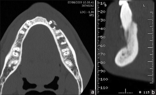
Malignant epithelioid hemangioendothelioma (MEH), or high-risk epithelioid hemangioendothelioma, is a low- to intermediate-grade vascular malignancy. A few cases of MEH have been documented in the head and neck region, including the neck, thyroid gland, larynx and scalp. MEHs are extremely rare in the oral cavity. Only 31 cases of MEH in the oral cavity were described in English literature between 1975 and 2014. Further, only eleven cases were referred to MEH of the maxillary or mandibular gingiva. No gingival MEH metastases have been described in literature. We report a literature review and a case of MEH with a metastatic occurrence 4 years after surgical excision.
Keywords: Malignant hemangioendothelioma; mandibular gingiva; vascular tumor.
Figures


Similar articles
-
Epithelioid Hemangioendothelioma of Mandibular Gingiva: A Challenging Diagnosis.J Clin Exp Dent. 2024 Sep 1;16(9):e1151-e1156. doi: 10.4317/jced.61925. eCollection 2024 Sep. J Clin Exp Dent. 2024. PMID: 39399857 Free PMC article.
-
Malignant epithelioid hemangioendothelioma of the lip: a case report and comprehensive literature review.J Oral Maxillofac Surg. 2014 Apr;72(4):695-701. doi: 10.1016/j.joms.2013.09.027. Epub 2013 Nov 20. J Oral Maxillofac Surg. 2014. PMID: 24268966 Review.
-
Primary epithelioid angiosarcoma originating from the mandibular gingiva: a case report of an extremely rare oral lesion.World J Surg Oncol. 2020 Oct 3;18(1):260. doi: 10.1186/s12957-020-01999-1. World J Surg Oncol. 2020. PMID: 33010804 Free PMC article. Review.
-
Epithelioid Haemangioendothelioma of the mandibular gingiva: case report and literature review.Int J Surg Case Rep. 2015;14:194-8. doi: 10.1016/j.ijscr.2015.06.041. Epub 2015 Jul 31. Int J Surg Case Rep. 2015. PMID: 26298095 Free PMC article.
-
[Case report of epithelioid hemangioendothelioma of the frontal region metastatic to the parotid gland].Laryngorhinootologie. 1998 Dec;77(12):728-31. doi: 10.1055/s-2007-997232. Laryngorhinootologie. 1998. PMID: 10036678 Review. German.
Cited by
-
Tonsillar Epithelioid Haemangioendotelioma: Description of a Rare Clinical Case.Indian J Otolaryngol Head Neck Surg. 2022 Dec;74(Suppl 3):5766-5768. doi: 10.1007/s12070-021-02386-2. Epub 2021 Jan 22. Indian J Otolaryngol Head Neck Surg. 2022. PMID: 36742620 Free PMC article.
-
Epithelioid Hemangioendothelioma of Mandibular Gingiva: A Challenging Diagnosis.J Clin Exp Dent. 2024 Sep 1;16(9):e1151-e1156. doi: 10.4317/jced.61925. eCollection 2024 Sep. J Clin Exp Dent. 2024. PMID: 39399857 Free PMC article.
-
Primary Intranodal Epithelioid Haemangioendothelioma in the Submandibular Region: A Case Report.Indian J Otolaryngol Head Neck Surg. 2024 Oct;76(5):3956-3961. doi: 10.1007/s12070-024-04752-2. Epub 2024 Jun 6. Indian J Otolaryngol Head Neck Surg. 2024. PMID: 39376453 Free PMC article.
-
Malignant Vascular Tumors of the Head and Neck-Which Type of Therapy Works Best?Cancers (Basel). 2021 Dec 9;13(24):6201. doi: 10.3390/cancers13246201. Cancers (Basel). 2021. PMID: 34944821 Free PMC article. Review.
-
Maxillary epithelioid hemangioendothelioma: an especially rare malignant tumor mimicking periodontal disease.BMC Oral Health. 2020 Nov 6;20(1):309. doi: 10.1186/s12903-020-01291-4. BMC Oral Health. 2020. PMID: 33158420 Free PMC article.
References
-
- Wesley RK, Mintz SM, Wertheimer FW. Primary malignant hemangioendothelioma of the gingiva. Oral Surg Oral Med Oral Pathol. 1975;39:103–112. - PubMed
-
- Weiss SW, Enzinger FM. Epithelioid hemangioendothelioma: A vascular tumor often mistaken for a carcinoma. Cancer. 1982;50:970–81. - PubMed
-
- Fletcher CD, Bridge JA, Hogendoorn P, Mertens F. 4 edition. Vol. 5. Lyon: IARC; 2013. WHO Classification of Tumours of Soft Tissue and Bone. World Health Organization.
-
- Makhlouf HR, Ishak KG, Goodman ZD. Epithelioid hemangioendothelioma of the liver: A clinicopathologic study of 137 cases. Cancer. 1999;85:562–82. - PubMed
-
- Mentzel T, Beham A, Calonje E, Katenkamp D, Fletcher CD. Epithelioid hemangioendothelioma of skin and soft tissues: Clinicopathologic and immunohistochemical study of 30 cases. Am J Surg Pathol. 1997;21:363–74. - PubMed
Publication types
LinkOut - more resources
Full Text Sources
Other Literature Sources
