. 2016 Feb 18;9(2):e1139252.
doi: 10.1080/19420889.2016.1139252. eCollection 2016 Mar-Apr.

Smaug variants in neural and non-neuronal cells

Affiliations

Smaug variants in neural and non-neuronal cells

Ana Julia Fernández-Alvarez et al. Commun Integr Biol. .

Abstract

Mammalian Smaug1/Samd4a is an mRNA regulator involved in synapse plasticity and additional non-neuronal functions. Here we analyzed the expression of Smaug1/Samd4a variants and Smaug2/Samd4b in primary hippocampal neurons and non-neuronal cell lines. We found that multiple Smaug proteins are present in several mammalian cell lines, including a canonical full length Smaug1, a Smaug1 variant that lacks the third exon, termed ΔEIII, and Smaug2, the product of a highly homologous gene. These three major isoforms are expressed differentially along neuron development and form cytosolic bodies when transfected in cell lines. By using luciferase reporters, we found that the ΔEIII isoform, which lacks 10 amino acids in the sterile α motif involved in RNA binding, shows a RNA-binding capacity and repressor activity comparable to that of the full length Smaug1. These observations are an important groundwork for molecular studies of the Smaug post-transcriptional pathway, which is relevant to neuron development, mitochondrial function and muscle physiology in health and disease.

Keywords: mRNA silencing; muscular dystrophy; processing bodies; stress granules; synaptogenesis; translation.


Figures

Figure 1.
Figure 1.
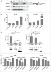
Smaug1 and Smaug2 variants in neurons and cell lines. (A) Schematic representation of full length (FL, NM_015589) and ΔEIII (NM_001161576) Smaug1. The ΔEIII lacks 10 amino acids in the SAM domain, which binds RNA. (B) RT-PCR of Smaug1 splicing variants. The presence of the full length and ΔEIII mRNAs was analyzed in neurons at 4, 8, 12 and 14 DIV by RT-PCR using isoform-specific primers (Material and Methods). Arrows indicate the position of each primer. C-, negative control (RNA sample with no reverse transcription). (C) Quantitative RT-PCR for total Smaug1 isoforms or Smaug2 in hippocampal neurons cultured during 8 or 14 d in vitro (DIV) was performed using the oligonucleotides indicated in Material and Methods. Results are expressed relative to β-actin mRNA levels. Both Smaug1 and Smaug2 transcripts accumulate during synaptogenesis in vitro. Error bars, s.e.m. *** p<0.001, Student's t-Test.
Figure 2.
Figure 2.
(A) The expression of Smaug1 variants and of Smaug2 was analyzed in the following cell lines: U2OS (U2), COS7 (C7), HEK293T (HK), SH-SY5Y (SH). A plasmid containing the complete Smaug1 or Smaug2 cDNA sequence was used in each case as a positive control (C+); C-, negative control (RNA sample with no reverse transcription). β-actin was analyzed for comparison. All cell lines express both Smaug1 isoforms and Smaug2. (B) Quantification of full length, ΔEIII, total Smaug1 or Smaug2 mRNAs in U2OS and HEK293T cells. Results are expressed as absolute cDNA in pg/µl, calculated using a standard curve obtained with plasmids containing the respective cDNAs as templates. Error bars, s.e.m. Statistical significance from Smaug2 relative to full length, ΔEIII and Smaug1 according to one-way ANOVA, Bonferroni post-test was *** p<0.0001. (C, D) Full length and ΔEIII Smaug1 bind and repress SRE-luciferase containing reporters. (C) The indicated Smaug1 constructs fused to a SBP-tag were co-transfected with firefly luciferase reporters carrying or not SREs (Materials and Methods). A plasmid encoding SBP-MBP was used as a negative control. The ratio in arbitrary units of luciferase mRNA in the pull down relative to that in total extracts is plotted. Western blot of pull down (PD) fractions or inputs (IN) and PD fractions are shown. The two Smaug1 variants display comparable binding activity. Error bars, s.e.m. Statistical significance according to one-way ANOVA, Bonferroni post-test was as follows: * p<0.05, ns, non-significant. (D), The indicated Smaug1 constructs fused to ECFP or V5 were co-transfected in BHK or U2OS cells together with Firefly luciferase reporters carrying wild type or mutant SRE motifs and a Renilla luciferase cDNA as a reference. Plasmids encoding V5 or ECFP were used as a control (Materials and Methods). The ratio of normalized luciferase wt SRE/mut SRE was plotted. Four representative experiments out of 6 are depicted. The two Smaug1 variants display comparable repressor activity whereas a construct lacking the SAM (ΔSAM) shows no effect. Error bars, s.e.m. Statistical significance relative to vector or ΔSAM according to one-way ANOVA, Bonferroni post-test was as follows: *** p<0.0001, ** p<0.001 and * p<0.05, respectively. No significant differences were seen between FL Smaug1 and ΔEIII and between the vector and ΔSAM.
Figure 3.
Figure 3.
Smaug1 and Smaug2 variants form cytosolic bodies. (A) U2OS cells were transfected with constructs for Smaug1, ΔEIII and Smaug2 fused to the indicated tags. The percentage of transfected cells with foci is shown in each case. (B) Smaug1, ΔEIII and Smaug2 constructs were co-transfected in pairs as indicated. The ΔEIII bodies colocalize almost completely with the bodies formed by the full length isoform. Smaug1 and Smaug2, as well as Smaug2 and ΔEIII also colocalize completely (for quantification see the main text). (C) The indicated constructs were co-transfected and endogenous XRN1 was stained with a specific antibody. XRN1 is present in foci (likely PBs), which are largely excluded from the Smaug1/2 bodies. Percentage of Smaug1/2 foci containing endogenous XRN1 is indicated for each construct.

References

    1. Thomas MG, Loschi M, Desbats MA, Boccaccio GL. RNA granules: the good, the bad and the ugly. Cell Signal 2011; 23:324-34; PMID:20813183; http://dx.doi.org/ 10.1016/j.cellsig.2010.08.011 - DOI - PMC - PubMed
    1. Baez MV, Luchelli L, Maschi D, Habif M, Pascual M, Thomas MG, Boccaccio GL. Smaug1 mRNA-silencing foci respond to NMDA and modulate synapse formation. J Cell Biol 2011; 195:1141-57; PMID:22201125; http://dx.doi.org/ 10.1083/jcb.201108159 - DOI - PMC - PubMed
    1. Luchelli L, Thomas MG, Boccaccio GL. Synaptic control of mRNA translation by reversible assembly of XRN1 bodies. J Cell Sci 2015; 128:1542-54; PMID:25736288; http://dx.doi.org/ 10.1242/jcs.163295 - DOI - PubMed
    1. Pascual ML, Luchelli L, Habif M, Boccaccio GL. Synaptic activity regulated mRNA-silencing foci for the fine tuning of local protein synthesis at the synapse. Commun Integr Biol 2012; 5:388-92; PMID:23060966; http://dx.doi.org/ 10.4161/cib.20257 - DOI - PMC - PubMed
    1. Chartier A, Klein P, Pierson S, Barbezier N, Gidaro T, Casas F, Carberry S, Dowling P, Maynadier L, Bellec M, et al.. Mitochondrial dysfunction reveals the role of mRNA poly(A) tail regulation in oculopharyngeal muscular dystrophy pathogenesis. PLoS Genet 2015; 11:e1005092; PMID:25816335; http://dx.doi.org/ 10.1371/journal.pgen.1005092 - DOI - PMC - PubMed

LinkOut - more resources