. 2016 Jan-Mar;11(1):70-7.
doi: 10.4103/2008-322X.180695.

Histological and Electrophysiological Changes in the Retinal Pigment Epithelium after Injection of Sodium Iodate in the Orbital Venus Plexus of Pigmented Rats

Affiliations

Histological and Electrophysiological Changes in the Retinal Pigment Epithelium after Injection of Sodium Iodate in the Orbital Venus Plexus of Pigmented Rats

Hamid Aboutaleb Kadkhodaeian et al. J Ophthalmic Vis Res. 2016 Jan-Mar.

Abstract

Purpose: To characterize histopathologic and electroretinographic (ERG) changes in the retina of pigmented rats injected with sodium iodate in order to establish a model of retinal degeneration for future cell therapy studies.

Methods: In 50 male pigmented rats weighing 250-300 grams, NaIO3 was injected into the left orbital venous plexus at 40 and 60 mg/kg doses (25 eyes in each group). Fourteen rats received phosphate buffered saline (PBS) injection in their left orbital plexus and were considered as the sham-control group. Histopathologic and ERG studies were performed at baseline and on days 1, 7, 14 and 28 after the injections.

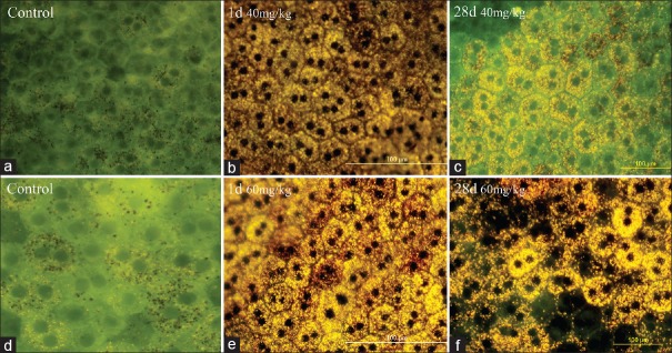
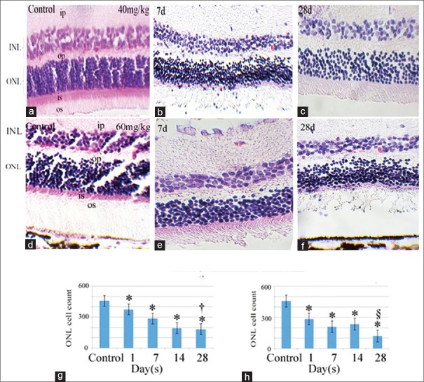
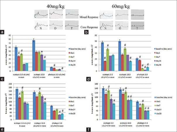
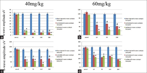
Results: Progressive retinal pigment epithelial (RPE) changes were observed from the first day of injection in both the 40 and 60 mg/kg study groups in a dose dependent manner. These changes manifested as loss of melanin pigment and accumulation of lipofuscin in RPE cells with subsequent cell death and patchy loss of RPE cells (in flat mounts), as well as thinning of the outer nuclear layer and later the inner nuclear layer in the succeeding days. ERG showed a progressive and significant decrease in a- and b- wave amplitudes in both case groups relative to baseline values and the controls (P < 0.05).

Conclusion: NaIO3 injection into the retrobulbar venous plexus of pigmented rats can result in significant and progressive damage to the RPE and subsequently to the neuroretina of the injected eye, and may serve as a model of retinal degeneration.

Keywords: Pigmented Rat; Retinal Pigment Epithelium; Retro-orbital Sinus; Sodium Iodate.


Figures

Figure 1
Figure 1
Whole flat-mount images of the RPE layer by fluorescence microscope. Images a and d show control specimens before sodium iodate injection. b and e represent specimens prepared one day (24 hours) after injection of 40 and 60 mg/kg retro-bulbar sodium iodate, respectively. Yellow-brown pigment granules in RPE cells may be the result of accumulation of lipofuscin secondary to sodium iodate injection. c andfrepresent specimens prepared 28 days after injection of 40 and 60 mg/kg of retrobulbar sodium iodate, respectively. Patchy disappearance of RPE cells is evident in specimens after 60 mg/kg injection. (Scale bar: 100 µm).
Figure 2
Figure 2
Histological cross section of the retina stained with hematoxylin and eosin. The upper panels (b and c) show specimens prepared 7 and 28 days after 40 mg/kg injection of retrobulbar sodium iodate. Lower panels (e and f) show specimens 7 and 28 days after 60 mg/kg of sodium iodate injection, respectively. a and d represent sections from normal retinas before any injection. Histograms (g and h, 40 and 60 mg/kg of sodium iodate, respectively) represent the morphometric evaluation of the outer nuclear layer (ONL cell count: Image J) in the control group and 1, 7, 14 and 28 days after injection. *This sign indicates a significant difference in ONL cell count between the control sections and sections of treated eyes from other time points. Indicates significantly lower ONL cell count in 28 days than the other time points except that of 14 days in 40 mg/kg group. § Indicates significantly lower ONL cell count than the other time points in 60 mg/kg group. OS, outer segment of photoreceptors; ONL, outer nuclear layer; OP, outer plexiform layer; INL, inner nuclear layer; IP, inner plexiform layer; Scale bar, A, B and D = 120 µm; C, E andF= 60 µm.
Figure 3
Figure 3
a and b show representative electroretinogram waveforms of the scotopic maximum combined and photopic responses, respectively. N: Before NaIO3 injection, O: One day and T: 28 days after sodium iodate administration at doses of 40 and 60 mg/kg, respectively. c and d represent the histograms of mean amplitudes of the a-wave with 40 and 60 mg/kg sodium iodate, from day 0 (before injection) to 1, 7, 14 and 28 days after injection, repectively. e andfrepresent mean amplitudes of the b-wave with 40 and 60 mg/kg sodium iodate, from day 0 (before injection) to 1, 7, 14 and 28 days after injection, respectively. (*Indicates a significant reduction in mean amplitudes compared to mean amplitudes recorded before Sodium iodate injection).
Figure 4
Figure 4
Comparison of mean a-wave amplitudes (a and b) and b-wave amplitudes (c and d) of the scotopic maximal combined electroretinograms (3 cd.s/m2 intensity), for eyes from the sham group (blue columns), sodium iodate injection of the left or ipsilateral eyes (green columns) and the right or contralateral eyes (red columns), of the 40 mg/kg (a and c) and 60 mg/kg (b and d) dose groups, in consecutive time points from before injection to 1, 7, 14 and 28 days after injection, respectively. (*Asterisk indicates a significant difference between the sham treated and other time point values in treated animals (P< 0.05). (#Indicates a significant difference between the contralateral eyes or red columns and ipsilateral eyes or green columns,P< 0.05).

Similar articles

Cited by

References

    1. Guymer R, Luthert P, Bird A. Changes in Bruch's membrane and related structures with age. Prog Retin Eye Res. 1999;18:59–90. - PubMed
    1. Bok D. The retinal pigment epithelium: A versatile partner in vision. J Cell Sci Suppl. 1993;17:189–195. - PubMed
    1. Marmor MF. Control of subretinal fluid: Experimental and clinical studies. Eye (Lond) 1990;4(Pt 2):340–344. - PubMed
    1. Gehrs KM, Anderson DH, Johnson LV, Hageman GS. Age-related macular degeneration – Emerging pathogenetic and therapeutic concepts. Ann Med. 2006;38:450–471. - PMC - PubMed
    1. Bird AC. Bruch's membrane change with age. Br J Ophthalmol. 1992;76:166–168. - PMC - PubMed