

Vascular access for extracorporeal life support: tips and tricks
- PMID: 27195133
- PMCID: PMC4856850
- DOI: 10.21037/jtd.2016.04.42
Vascular access for extracorporeal life support: tips and tricks
Abstract
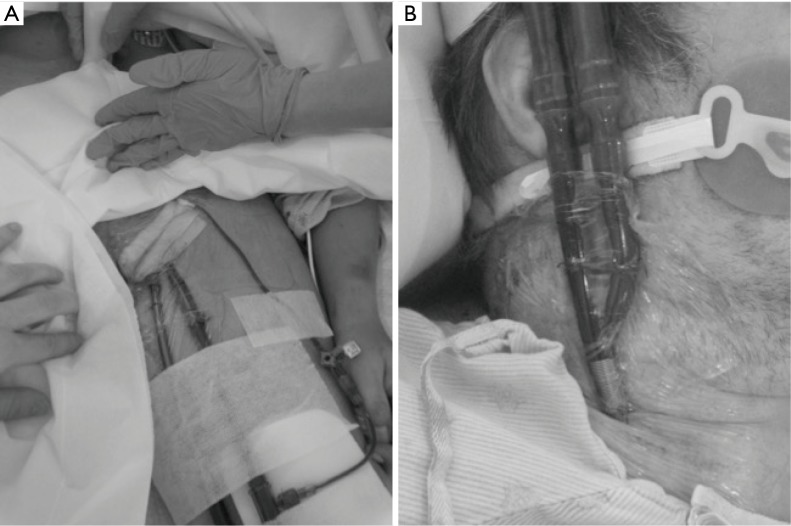
In thoracic surgery, extracorporeal life support (ECLS) techniques are performed to (I) provide a short to mid term extracorporeal mechanical support; (II) realize the gas exchanges; and (III)-depending the configuration of the circuit-substitute the failed heart function. The objective of this review is to describe the rational of the different ECLS techniques used in thoracic surgery and lung transplantation (LTx) with a specific attention to the vascular access. Venovenous extracorporeal membrane oxygenation (VV ECMO) is the most common ECLS technique used in thoracic surgery and represents the best strategy to support the lung function. VV ECMO needs peripheral vascular access. The selection between his double-site or single-site configuration should be decided according the level of O2 requirements, the nosological context, and the interest to perform an ECLS ambulatory strategy. Venoarterial (VA) ECMO uses peripheral and/or central cannulation sites. Central VA ECMO is mainly used in LTx instead a conventional cardiopulmonary bypass (CPB) to decrease the risk of hemorrhagic issues and the rate of primary graft dysfunction (PGD). Peripheral VA ECMO is traditionally realized in a femoro-femoral configuration. Femoro-femoral VA ECMO allows a cardiocirculatory support but does not provide an appropriate oxygenation of the brain and the heart. The isolated hypercapnic failure is currently supported by extracorporeal CO2 removal (ECCO2R) devices inserted in jugular or subclavian veins. The interest of the Novalung (Novalung GmbH, Hechingen, Germany) persists due to his central configuration indicated to bridge to LTx patients suffering from pulmonary hypertension. The increasing panel of ECLS technologies available in thoracic surgery is the results of a century of clinical practices, engineering progress, and improvements of physiological knowledges. The selection of the ECLS technique-and therefore the vascular access to implant the device-for a given nosological context trends to be defined according an evidence-based medicine.
Keywords: Extracorporeal life support/extracorporeal membrane oxygenation (ECLS/ECMO); ICU; acute respiratory distress syndrome (ARDS); lung transplantation (LTx); thoracic surgery.
Conflict of interest statement
Figures


References
-
- Bartlett RH, Gazzaniga AB, Jefferies MR, et al. Extracorporeal membrane oxygenation cardiopulmonary support in infancy. Trans Am Soc Artif Intern Organs 1976;22:80-93. - PubMed
-
- Kolobow T. Gas exchange with membrane lungs. In: Gille JP, editor. Neonatal and adult respiratory failure: mechanisms and treatment. Paris: Elsevier, 1989:89-96.
-
- Gattinoni L, Kolobow T, Damia G, et al. Extracorporeal carbon dioxide removal (ECCO2R): a new form of respiratory assistance. Int J Artif Organs 1979;2:183-5. - PubMed
Publication types
LinkOut - more resources
Full Text Sources
Other Literature Sources
Miscellaneous