Review
. 2016:2016:6137541.
doi: 10.1155/2016/6137541. Epub 2016 Apr 18.

Tricellular Tight Junctions in the Inner Ear

Affiliations
Review

Tricellular Tight Junctions in the Inner Ear

Shin-Ichiro Kitajiri et al. Biomed Res Int. 2016.

Abstract

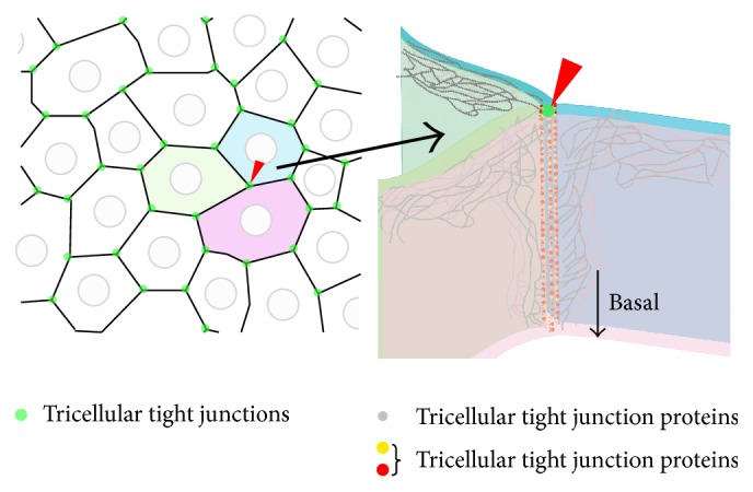
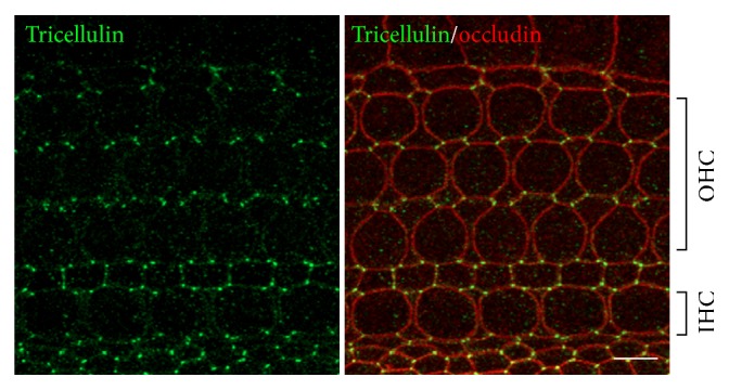
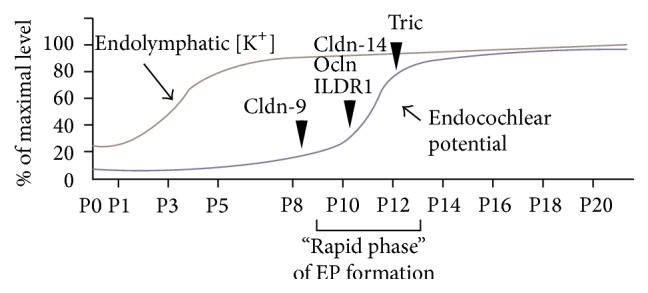
Tight junctions (TJs) are structures that seal the space between the epithelial cell sheets. In the inner ear, the barrier function of TJs is indispensable for the separation of the endolymphatic and perilymphatic spaces, which is essential for the generation and maintenance of the endocochlear potential (EP). TJs are formed by the intercellular binding of membrane proteins, known as claudins, and mutations in these proteins cause deafness in humans and mice. Within the epithelial cell sheet, however, a bound structure is present at the site where the corners of three cells meet (tricellular tight junctions (tTJs)), and the maintenance of the barrier function at this location cannot be explained by the claudins alone. Tricellulin and the angulin family of proteins (angulin-1/LSR, angulin-2/ILDR1, and angulin-3/ILDR2) have been identified as tTJ-associated proteins. Tricellulin and ILDR1 are localized at the tTJ and alterations in these proteins have been reported to be involved in deafness. In this review, we will present the current state of knowledge for tTJs.


Figures

Figure 1
Figure 1
Schematic image of the tricellular tight junction (tTJ). The TJs between two cells (bicellular TJ, bTJ) function to seal off the intercellular space (black lines). However, the corner where three cells meet (green dots) cannot be occluded by bTJ proteins alone and a central “tube” may exist. tTJ proteins (red dots and yellow dots) are localized at this site and play a role in providing a barrier.
Figure 2
Figure 2
Localization of tricellulin and occludin in the organ of Corti. Double immunofluorescence microscopy of the organ of Corti of postnatal day 3 C57BL/6 mice using anti-tricellulin pAb (green) and anti-occludin mAb (red). Tricellulin is localized at tricellular contacts where three cells meet. Occludin, a tight junction-associated membrane protein, distributed to cell-cell contacts not only at tTJ but also at bicellular TJ. OHC: outer hair cell; IHC: inner hair cell. Bars, 5 μm.
Figure 3
Figure 3
Time course of EP and endolymphatic [K+] elevation (modified from [–41]) and hair cell degeneration in mice with mutations in TJ proteins. The arrowheads indicate the starting time for hair cell degeneration in each of the mutant mice models: cldn-9: claudin-9 mutant mice [18]; cldn-14: claudin-14 knockout mice [17]; ocln: occludin deficient mice [33]; ILDR1: ILDR1 null mice [34, 35]; Tric: tricellulin knockin mice [29] and knockout mice [36].

References

    1. Richardson G. P., de Monvel J. B., Petit C. How the genetics of deafness illuminates auditory physiology. Annual Review of Physiology. 2011;73:311–334. doi: 10.1146/annurev-physiol-012110-142228. - DOI - PubMed
    1. Ferrary E., Sterkers O. Mechanisms of endolymph secretion. Kidney International. 1998;65:S98–S103. - PubMed
    1. Nunes F. D., Lopez L. N., Lin H. W., et al. Distinct subdomain organization and molecular composition of a tight junction with adherens junction features. Journal of Cell Science. 2006;119(part 23):4819–4827. doi: 10.1242/jcs.03233. - DOI - PubMed
    1. Steed E., Balda M. S., Matter K. Dynamics and functions of tight junctions. Trends in Cell Biology. 2010;20(3):142–149. doi: 10.1016/j.tcb.2009.12.002. - DOI - PubMed
    1. Angelow S., Ahlstrom R., Yu A. S. L. Biology of claudins. The American Journal of Physiology—Renal Physiology. 2008;295(4):F867–F876. doi: 10.1152/ajprenal.90264.2008. - DOI - PMC - PubMed

Substances