. 2016 May 19;11(5):e0155824.
doi: 10.1371/journal.pone.0155824. eCollection 2016.

Rescuing Perishable Neuroanatomical Information from a Threatened Biodiversity Hotspot: Remote Field Methods for Brain Tissue Preservation Validated by Cytoarchitectonic Analysis, Immunohistochemistry, and X-Ray Microcomputed Tomography

Affiliations

Rescuing Perishable Neuroanatomical Information from a Threatened Biodiversity Hotspot: Remote Field Methods for Brain Tissue Preservation Validated by Cytoarchitectonic Analysis, Immunohistochemistry, and X-Ray Microcomputed Tomography

Daniel F Hughes et al. PLoS One. .

Abstract

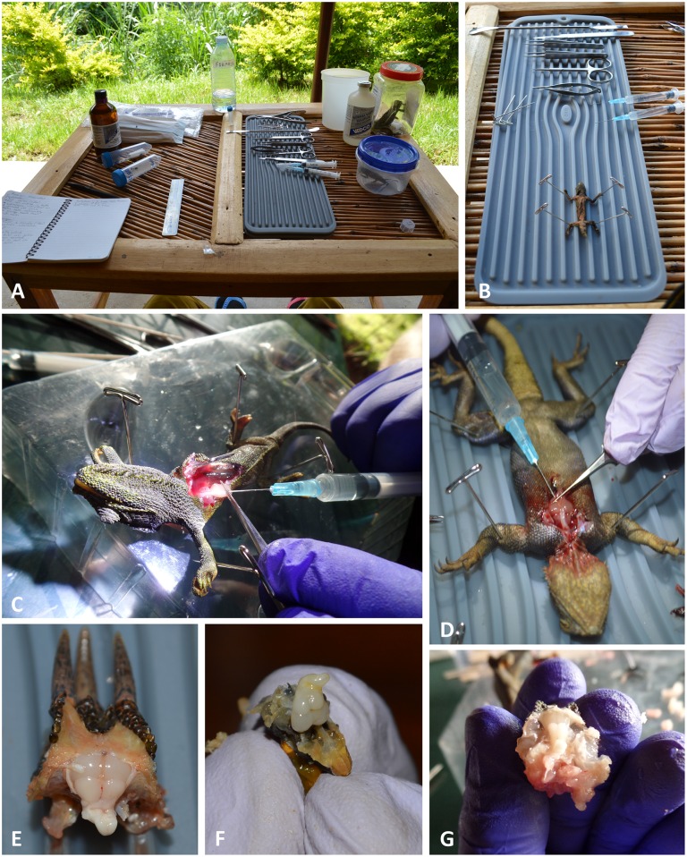
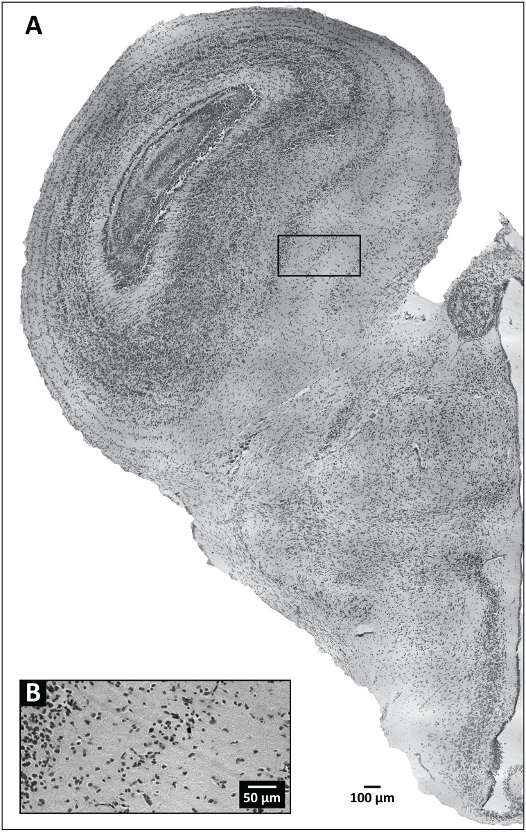
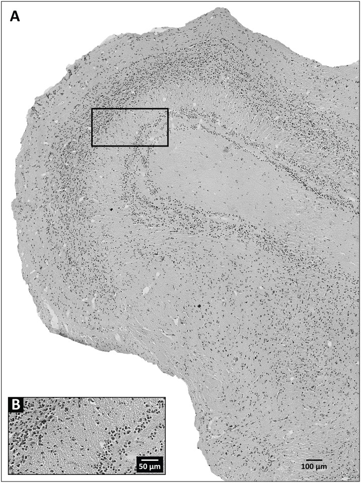
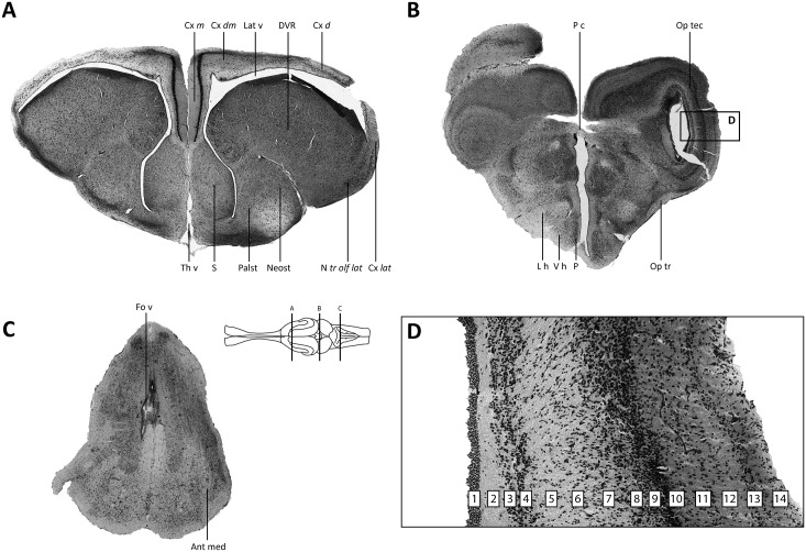
Biodiversity hotspots, which harbor more endemic species than elsewhere on Earth, are increasingly threatened. There is a need to accelerate collection efforts in these regions before threatened or endangered species become extinct. The diverse geographical, ecological, genetic, morphological, and behavioral data generated from the on-site collection of an individual specimen are useful for many scientific purposes. However, traditional methods for specimen preparation in the field do not permit researchers to retrieve neuroanatomical data, disregarding potentially useful data for increasing our understanding of brain diversity. These data have helped clarify brain evolution, deciphered relationships between structure and function, and revealed constraints and selective pressures that provide context about the evolution of complex behavior. Here, we report our field-testing of two commonly used laboratory-based techniques for brain preservation while on a collecting expedition in the Congo Basin and Albertine Rift, two poorly known regions associated with the Eastern Afromontane biodiversity hotspot. First, we found that transcardial perfusion fixation and long-term brain storage, conducted in remote field conditions with no access to cold storage laboratory equipment, had no observable impact on cytoarchitectural features of lizard brain tissue when compared to lizard brain tissue processed under laboratory conditions. Second, field-perfused brain tissue subjected to prolonged post-fixation remained readily compatible with subsequent immunohistochemical detection of neural antigens, with immunostaining that was comparable to that of laboratory-perfused brain tissue. Third, immersion-fixation of lizard brains, prepared under identical environmental conditions, was readily compatible with subsequent iodine-enhanced X-ray microcomputed tomography, which facilitated the non-destructive imaging of the intact brain within its skull. In summary, we have validated multiple approaches to preserving intact lizard brains in remote field conditions with limited access to supplies and a high degree of environmental exposure. This protocol should serve as a malleable framework for researchers attempting to rescue perishable and irreplaceable morphological and molecular data from regions of disappearing biodiversity. Our approach can be harnessed to extend the numbers of species being actively studied by the neuroscience community, by reducing some of the difficulty associated with acquiring brains of animal species that are not readily available in captivity.


Conflict of interest statement

Competing Interests: The authors have declared that no competing interests exist.

Figures

Fig 1
Fig 1. Images of field-based perfusion technique.
The field laboratory setup (A); pinned lizard on silicone mat prior to opening of the thoracic cavity (B); injections of solution through opening in apex of heart (C, D); partially dissected and exposed formaldehyde-fixed brains (E, F, G).
Fig 2
Fig 2. Representative tissue section at the level of the optic tectum, obtained from a field-perfused specimen of Trioceros johnstoni.
(A) Wide field image of a hemisphere from a section of the specimen. The black box outlines the area enlarged in (B), which provides details regarding the level of background staining and cellular labeling demonstrable by our Nissl-based staining procedure.
Fig 3
Fig 3. Representative tissue section at the level of the optic tectum, obtained from a laboratory-perfused specimen of Trioceros jacksonii.
(A) Wide field image of a hemisphere from a section of the specimen. The black box outlines the area enlarged in (B), which provides details regarding the level of background staining and cellular labeling demonstrable by our Nissl-based staining procedure.
Fig 4
Fig 4. Photomicrographs of Nissl-stained brain sections from an agamid lizard.
(A–C) Major brain regions are represented (A—forebrain; B—midbrain; C—hindbrain). (D) Detailed image of the optic tectum. The brain schematic was adapted from a drawing of a lizard rendered by artist Christiaan van Huijzen for Poster 2 of the poster book accompanying [40]. Delineation of major brain regions (A–C) generally follows [–49] with only cosmetic changes made to the abbreviation style. The laminar organization of the optic tectum (D) follows [50]. Abbreviations: Ant med—Anterior medulla; Cx d—Cortex dorsalis (dorsal cortex); Cx dm—Cortex dorsomedialis (dorsomedial cortex); Cx lat—Cortex lateralis (lateral cortex); Cx m–Cortex medialis (medial cortex); DVR—Dorsal Ventricular Ridge; Fo v—Fourth ventricle; L h—Lateral hypothalamus; Lat v—Lateral ventricle; Neost—Neostriatum; N tr olf lat–Nucleus tractus olfactori lateralis (nucleus of the lateral olfactory tract); Op tr—Optic tract; Op tec—Optic tectum; Palst—Paleostriatum; P—Periventricular hypothalamus; P c—Posterior commissure; S—septal nuclei; Th v—Third ventricle; V h—Ventral hypothalamus.
Fig 5
Fig 5. Heat map of scored semi-quantitative data for six qualitative variables from three independent observers.
Observers evaluated Nissl-stained tissue sections (n = 204) from laboratory and field treatments using solutions containing 4% formaldehyde. Each column indicated with a small number (1, 2, or 3) represents an observer. Columns are grouped according to the qualitative variable being rated: presence of blood in tissue (A); evenness of stain (B); integrity of tissue at center of section (C); integrity of tissue at edges of section (D); clarity of lamination patterns (E); visibility of cell areas and nuclei (F). Tissue sections prepared under laboratory conditions are positioned on top (n = 166) and those prepared under field conditions on bottom (n = 38). The color code for the scored data is shown above the heat map. The black box outlines denoted with a ‘-‘ or a ‘+’ indicate selected regions of tissue ratings (negative or positive, respectively) that were largely uniform across observers.
Fig 6
Fig 6. Comparison of immunohistochemical staining of brain tissue fixed under field and laboratory conditions.
(A, B). The images show tyrosine hydroxylase immunoreactivity (-ir) (TH; red) with DAPI fluorescent counterstain (blue) for (A) Trioceros johnstoni fixed under field conditions, and (B) Rieppeleon kerstenii fixed under laboratory conditions. (C, D). The images show neuropeptide Y-ir (NPY; green), again with DAPI (blue) for (C) Rhampholeon boulengeri fixed under field conditions and (D) Rieppeleon kerstenii fixed under laboratory conditions (note that tissues in B and D are from the same animal). Both immunoreactive neurons (arrows) and neuronal extensions (small solid horizontal lines ending in hollow circles) are clearly visible, many of the latter being identifiable axons with varicosities. The ependymal cell layers lining the third ventricle (Th v) in A, C and D are indicated by arrowheads. The single-plane image in A rendered a portion of the image slightly out of focus (asterisk). Insets (a–d) show views of sections processed in the absence of the primary antibody. For inset b, the image has been brightened linearly so that the tissue section can be clearly seen in the photo. Scale bars (panel A, inset a) apply to all remaining panels and insets, respectively.
Fig 7
Fig 7. Diffusible iodine-based contrast-enhanced computed tomography (DiceCT) through the heads of two chameleon species.
(A) Parasagittal view of an adult male representative of Rhampholeon boulengeri; (B) parasagittal and (C) frontal views of an adult female representative of Trioceros johnstoni. These images illustrate the extraordinary diversity of soft anatomical structures that can be clearly visualized with our approach, including myelinated and unmyelinated components of the brain. Abbreviations for selected structures: ACC—M. accelerator linguae; BH—basihyoid; BS—brain stem; DVR—dorsal ventricular ridge; ENT—entoglossal process; HG—M. hyoglossus; ON—optic nerve; Op tec—Optic tectum; OTr—olfactory tract; OV—optic ventricle; RET—retina; SC—spinal cord; Th v—third ventricle; TP—tongue pad.

References

    1. Mora C, Tittensor DP, Adl S, Simpson AGB, Worm B. How many species are there on Earth and in the ocean? PLoS Biol. 2011; 9: e1001127 10.1371/journal.pbio.1001127 - DOI - PMC - PubMed
    1. Mittermeier RA, Turner WA, Larsen FW, Brooks TM, Gascon C. Global biodiversity conservation: The critical role of hotspots In: Zachos FE, Habel JC, editors. Biodiversity hotspots: Distribution and protection of conservation priority areas. Berlin: Springer-Verlag; 2011. p. 3–22.
    1. Myers N. Biodiversity hotspots revisited. Biosci. 2003; 53: 916–917.
    1. Hoffman M, Hilton-Taylor C, Angulo A, Böhm M, Brooks TM, Butchart SHM et al. The impact of conservation on the status of the world’s vertebrates. Science. 2010; 230: 1503–1509. - PubMed
    1. Iwaniuk AN. The importance of scientific collecting and natural history museums for comparative neuroanatomy. Ann N Y Acad Sci. 2011; 1225 (Suppl 1): E1–E19. - PubMed

Publication types

LinkOut - more resources