Review
. 2016 Aug;37(4):372-402.
doi: 10.1210/er.2016-1007. Epub 2016 May 19.

Estrogens, Neuroinflammation, and Neurodegeneration

Affiliations
Review

Estrogens, Neuroinflammation, and Neurodegeneration

Alessandro Villa et al. Endocr Rev. 2016 Aug.

Abstract

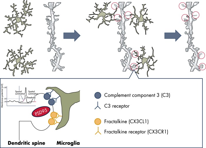
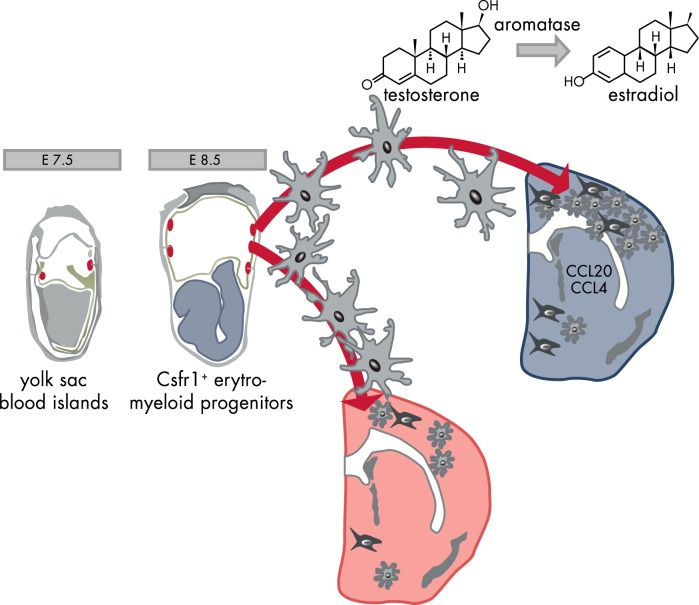
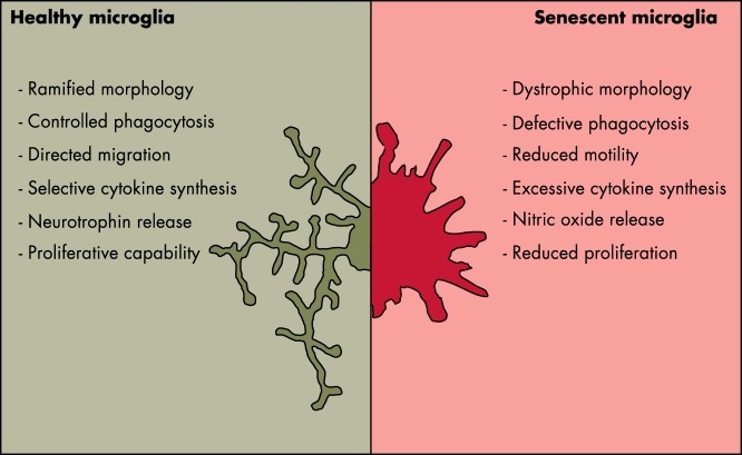
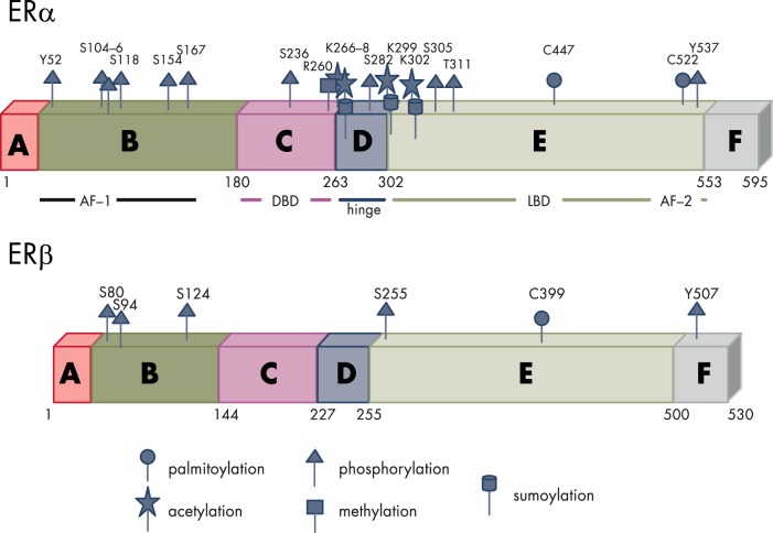
Inflammatory activation of microglia is a hallmark of several disorders of the central nervous system. In addition to protecting the brain against inflammatory insults, microglia are neuroprotective and play a significant role in maintaining neuronal connectivity, but the prolongation of an inflammatory status may limit the beneficial functions of these immune cells. The finding that estrogen receptors are present in monocyte-derived cells and that estrogens prevent and control the inflammatory response raise the question of the role that this sex steroid plays in the manifestation and progression of pathologies that have a clear sex difference in prevalence, such as multiple sclerosis, Parkinson's disease, and Alzheimer's disease. The present review aims to provide a critical review of the current literature on the actions of estrogen in microglia and on the involvement of estrogen receptors in the manifestation of selected neurological disorders. This current understanding highlights a research area that should be expanded to identify appropriate replacement therapies to slow the progression of such diseases.


Figures

Figure 1.
Figure 1.
Microglia are a dynamic mediator of synaptic development and homeostasis. Microglia in its surveying state senses the state of activity of neurons, is attracted to the dendritic spines through proteins of the complement and fractalkine, and participates in neuronal plasticity potentially through the release of proteases able to modulate the structure and functions of the synapses. Microglia possibly respond to the release of ATP, which may induce the shedding of lipid-rich vesicles that were reported to increase the frequency and amplitude of the excitatory postsynaptic potential.
Figure 2.
Figure 2.
Brain development: the sexually dimorphic activity of microglia. Primitive macrophages exit the yolk sac blood islands at the onset of circulation and colonize the neuroepithelium from E8.5 to give rise to microglia. The BBB starts to form from E13.5 and may isolate the developing brain from the contribution of fetal liver hematopoiesis. Embryonic microglia expand and colonize the whole CNS until adulthood. The high concentration of chemokines in the male brain facilitates microglia proliferation. E, embryonic day.
Figure 3.
Figure 3.
Morphological and functional elements point to alterations of microglia activity with age.
Figure 4.
Figure 4.
Structure and PTM sites of the nuclear ERs. Like all steroid receptors, the ERs belong to a family of hormone-modulated transcription factors characterized by the presence of six functional domains: A and B, the N-terminal domain that contains the activation function 1 (AF-1) enabling the interaction with coregulators also in the absence of the ligand. C, The highly conserved DBD, responsible for the recognition of specific DNA sequences (named estrogen responsive elements or EREs) through the two Zn fingers. D, The hinge region, a flexible domain that connects DBD with the ligand binding domain (LBD) able to influence intracellular trafficking and subcellular distribution. E, The LBD responsible for ligand recognition that contains the ligand-dependent activation function 2 (AF-2); the LBD, contributes to the dimerization interface of the receptor in concert with the DBD. F, The C-terminal domain that participates in the binding to ligands. Both ERs undergo a large number of regulatory PTMs exemplified in the figure (human ERα and murine ERβ).
Figure 5.
Figure 5.
Aging effects on circulating estrogens. The uterus weight as a biomarker of circulating estrogens shows that, in mice, the activity of the ovaries does not decrease with age; actually at 18 months, when mice are not cycling, the plasma content of this hormone is higher than in young, fertile animals. Ovx clearly decreases the circulating levels of the hormone, showing that organs other than ovaries give a minimal contribution to steroidogenesis. °: P < 0.05; °°°: P < 0.001; *: P < 0.05; n.d., not detected.
Figure 6.
Figure 6.
Estrogen and microglia functions. Estrogens regulate microglia inflammatory potential by interfering with the process of NF-κB activation (left) and by facilitating the transition to the stages where microglia exert neuroprotective functions (right), possibly including the maintenance and pruning of dysfunctional synapses (bottom).
Figure 7.
Figure 7.
Estrogen-dependent protective effects of microglia. Several biochemical processes promoted by microglia and regulated by estrogens protect neuronal functions: phagocytosis clears the debris and dysfunctional proteins (ie, β-amyloid) in the parenchyma; the production of antioxidant systems and enzymes (ie, the renin-angiotensin axis) limits the oxidative stress; the healing process is facilitated when damage-activated intracellular signaling pathways are activated; and synaptic maintenance participates in neuronal signaling. With aging, misfolded proteins, cell debris, and other inflammatory stimuli accumulate in the brain parenchyma, inducing a continuous stimulation of microglia that with senescence has a decreased phagocytic potential and ability to return to the surveying state. This initiates a vicious cycle with a progressive increase of the production of inflammatory products detrimental for neuronal health.

References

    1. Milligan ED, Watkins LR. Pathological and protective roles of glia in chronic pain. Nat Rev Neurosci. 2009;10(1):23–36. - PMC - PubMed
    1. Vezzani A, French J, Bartfai T, Baram TZ. The role of inflammation in epilepsy. Nat Rev Neurol. 2011;7(1):31–40. - PMC - PubMed
    1. Friedrich MJ. Research on psychiatric disorders targets inflammation. JAMA. 2014;312(5):474–476. - PubMed
    1. Perry VH, Holmes C. Microglial priming in neurodegenerative disease. Nat Rev Neurol. 2014;10(4):217–224. - PubMed
    1. Kettenmann H, Hanisch UK, Noda M, Verkhratsky A. Physiology of microglia. Physiol Rev. 2011;91(2):461–553. - PubMed

Publication types

MeSH terms