. 2016 May 19;11(5):e0155983.
doi: 10.1371/journal.pone.0155983. eCollection 2016.

Inhibition of β-Catenin to Overcome Endocrine Resistance in Tamoxifen-Resistant Breast Cancer Cell Line

Affiliations

Inhibition of β-Catenin to Overcome Endocrine Resistance in Tamoxifen-Resistant Breast Cancer Cell Line

Hye Sung Won et al. PLoS One. .

Abstract

Background: The β-catenin signaling is important in cell growth and differentiation and is frequently dysregulated in various cancers. The most well-known mechanism of endocrine resistance is cross-talk between the estrogen receptor (ER) and other growth factor signaling, such as phosphatidylinositol-3-kinase (PI3K)/Akt and the mammalian target of rapamycin (mTOR) signaling pathway. In the present study, we investigated whether β-catenin could be a potential target to overcome endocrine resistance in breast cancer.

Methods: We established tamoxifen-resistant (TamR) cell line via long-term exposure of MCF-7 breast cancer cells to gradually increasing concentrations of tamoxifen. The levels of protein expression and mRNA transcripts were determined using western blot analysis and real-time quantitative PCR. The transcriptional activity of β-catenin was measured using luciferase activity assay.

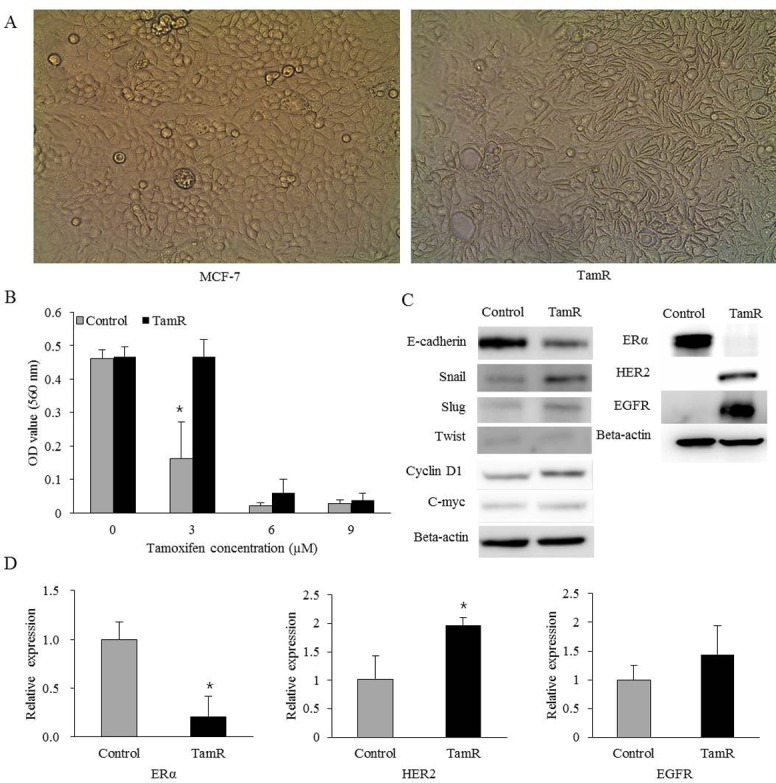
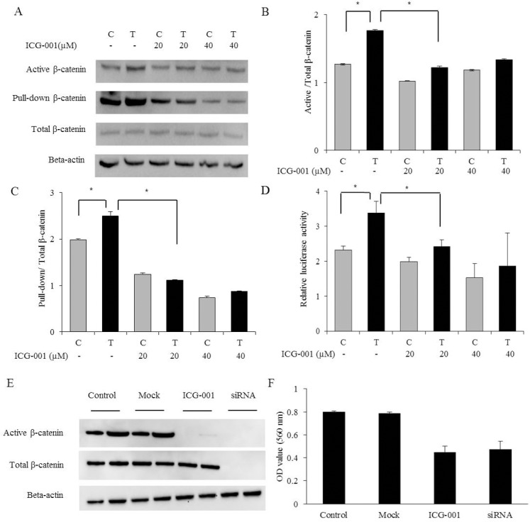
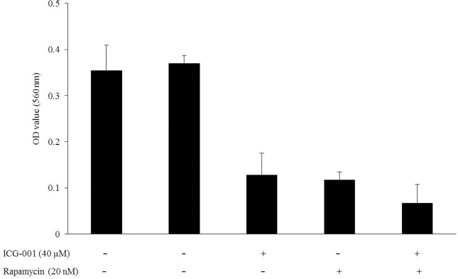
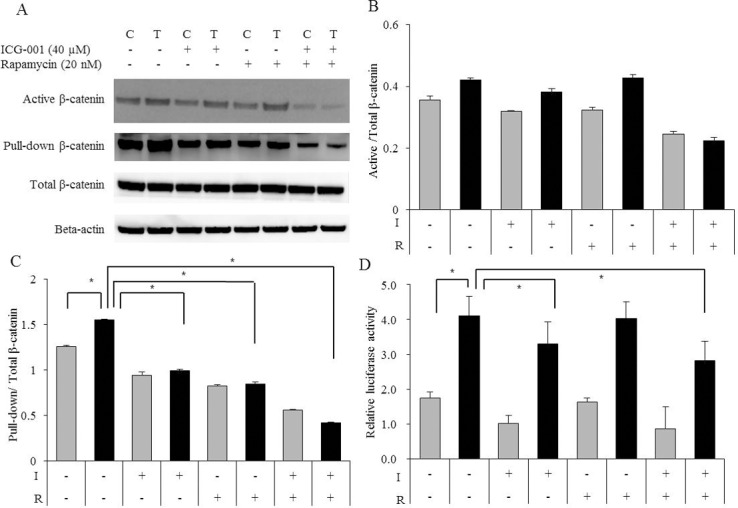
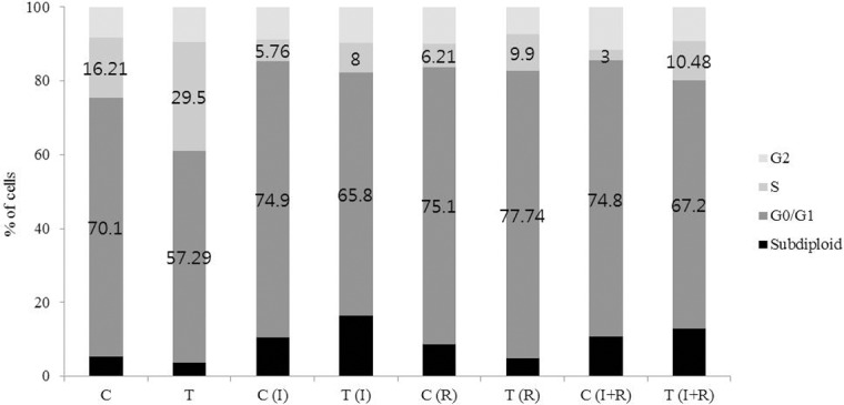
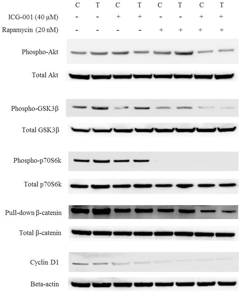
Results: TamR cells showed a mesenchymal phenotype, and exhibited a relatively decreased expression of ER and increased expression of human epidermal growth factor receptor 2 and the epidermal growth factor receptor. We confirmed that the expression and transcriptional activity of β-catenin were increased in TamR cells compared with control cells. The expression and transcriptional activity of β-catenin were inhibited by β-catenin small-molecule inhibitor, ICG-001 or β-catenin siRNA. The viability of TamR cells, which showed no change after treatment with tamoxifen, was reduced by ICG-001 or β-catenin siRNA. The combination of ICG-001 and mTOR inhibitor, rapamycin, yielded an additive effect on the inhibition of viability in TamR cells.

Conclusion: These results suggest that β-catenin plays a role in tamoxifen-resistant breast cancer, and the inhibition of β-catenin may be a potential target in tamoxifen-resistant breast cancer.


Conflict of interest statement

Competing Interests: The authors have declared that no competing interests exist.

Figures

Fig 1
Fig 1. Characteristics of tamoxifen-resistant breast cancer cell lines.
(A) TamR cells exhibited a more elongated, spindle-shaped morphology, whereas control cells exhibited a cuboidal shape (magnification 100×). (B) The viability of control cells decreased by 65.2% after 3 μM tamoxifen treatment, whereas the viability of TamR cells showed no change. (C) Western blot analysis of EMT-related markers, estrogen receptor alpha (ERα), human epidermal growth factor receptor 2 (HER2) and epidermal growth factor receptor (EGFR) in control and TamR cells. TamR cells showed decreased expression of E-cadherin and increased expression of Snail, Slug, and cyclin D1. TamR cells showed decreased expression of ERα and increased expression of HER2 and EGFR. (D) mRNA expression of ERα, HER2 and EGFR was assessed by RT-PCR. TamR cells exhibited a relatively decreased expression of ERα and increased expression of HER2 and EGFR. Error bars, mean ± standard deviation (SD). *, P < 0.05.
Fig 2
Fig 2. Protein expression and transcriptional activity of β-catenin and its inhibitory effects in TamR cells.
(A) Protein expression levels of β-catenin were measured by western blot analysis using a commercial primary antibody and pull-down assay. The expression of β-catenin was increased in TamR cells and was inhibited by ICG-001. (B) Quantification of band intensities was determined by densitometry analysis. The graphs showed the active β-catenin divided by total β-catenin. (C) Quantification of band intensities was determined by densitometry analysis. The graphs showed the pull-down β-catenin divided by total β-catenin. (D) The transcriptional activity of β-catenin was assessed using luciferase reporter assay. The transcriptional activity of β-catenin was increased in TamR cells and was inhibited by ICG-001. (E) The protein expression of β-catenin was also inhibited by β-catenin siRNA. (F) Cell viability assay of TamR cells was assessed after treatment with ICG-001 (40 μM) and β-catenin siRNA for 24 hours. The viability of TamR cells was decreased by ICG-001 and β-catenin siRNA. The experiment was repeated three times. Error bars, mean ± SD. *, P < 0.05. C, control cells. T, TamR cells.
Fig 3
Fig 3. Combinatory effect of ICG-001 and rapamycin in TamR cells.
Cell viability assay of TamR cells after treatment with ICG-001 (40 μM) and rapamycin (20 nM) for 24 hours was performed. The viability of TamR cells, which had showed resistance to tamoxifen (3 μM), was decreased by ICG-001 and rapamycin. All media except first lane were treated with tamoxifen (3 μM).
Fig 4
Fig 4. Alteration of β-catenin after treatment with ICG-001 and rapamycin.
(A) The expression of β-catenin was decreased by ICG-001 and rapamycin. It was decreased to a greater extent by the combination of ICG-001 and rapamycin. (B) Quantification of band intensities was determined by densitometry analysis. The graphs showed the active β-catenin divided by total β-catenin. (C) Quantification of band intensities was determined by densitometry analysis. The graphs showed the pull-down β-catenin divided by total β-catenin. (D) The transcriptional activity of β-catenin reduced to the greatest extent by the combination of ICG-001 and rapamycin. The experiment was repeated three times. Error bars, mean ± SD. *, P < 0.05. I, ICG-001 (40 μM). R, Rapamycin (20 nM). C, control cells. T, TamR cells.
Fig 5
Fig 5. Cell cycle analysis of control and TamR cells with ICG-001 and rapamycin.
Flow cytometric analysis of cell cycle was performed in control and TamR cells after treatment with ICG-001 (40 μM) and rapamycin (20 nM) for 24 hours. TamR cells showed the accelerated G1 to S phase transition. ICG-001 increased the fraction of apoptotic cells and rapamycin increased the fraction of G0/G1-arrested cells. I, ICG-001 (40 μM). R, Rapamycin (20 nM). C, control cells. T, TamR cells.
Fig 6
Fig 6. Alteration of PI3K/Akt/mTOR and β-catenin pathway- related proteins after treatment with ICG-001 and rapamycin.

References

    1. Eroles P, Bosch A, Perez-Fidalgo JA, Lluch A. Molecular biology in breast cancer: intrinsic subtypes and signaling pathways. Cancer Treat Rev. 2012;38(6):698–707. 10.1016/j.ctrv.2011.11.005 - DOI - PubMed
    1. Ziauddin MF, Hua D, Tang SC. Emerging strategies to overcome resistance to endocrine therapy for breast cancer. Cancer Metastasis Rev. 2014;33(2–3):791–807. 10.1007/s10555-014-9504-6 - DOI - PubMed
    1. Ring A, Dowsett M. Mechanisms of tamoxifen resistance. Endocr Relat Cancer. 2004;11(4):643–58. - PubMed
    1. Musgrove EA, Sutherland RL. Biological determinants of endocrine resistance in breast cancer. Nat Rev Cancer. 2009;9(9):631–43. 10.1038/nrc2713 - DOI - PubMed
    1. Osborne CK, Schiff R. Mechanisms of endocrine resistance in breast cancer. Annu Rev Med. 2011;62:233–47. 10.1146/annurev-med-070909-182917 - DOI - PMC - PubMed

Publication types

MeSH terms