. 2016 Jun 1;76(11):3376-86.
doi: 10.1158/0008-5472.CAN-15-2654. Epub 2016 Apr 11.

Ly6E/K Signaling to TGFβ Promotes Breast Cancer Progression, Immune Escape, and Drug Resistance

Affiliations

Ly6E/K Signaling to TGFβ Promotes Breast Cancer Progression, Immune Escape, and Drug Resistance

Midrar AlHossiny et al. Cancer Res. .

Abstract

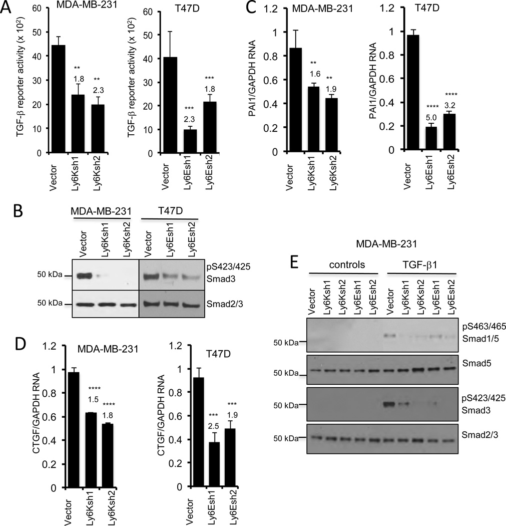
Stem cell antigen Sca-1 is implicated in murine cancer stem cell biology and breast cancer models, but the role of its human homologs Ly6K and Ly6E in breast cancer are not established. Here we report increased expression of Ly6K/E in human breast cancer specimens correlates with poor overall survival, with an additional specific role for Ly6E in poor therapeutic outcomes. Increased expression of Ly6K/E also correlated with increased expression of the immune checkpoint molecules PDL1 and CTLA4, increased tumor-infiltrating T regulatory cells, and decreased natural killer (NK) cell activation. Mechanistically, Ly6K/E was required for TGFβ signaling and proliferation in breast cancer cells, where they contributed to phosphorylation of Smad1/5 and Smad2/3. Furthermore, Ly6K/E promoted cytokine-induced PDL1 expression and activation and binding of NK cells to cancer cells. Finally, we found that Ly6K/E promoted drug resistance and facilitated immune escape in this setting. Overall, our results establish a pivotal role for a Ly6K/E signaling axis involving TGFβ in breast cancer pathophysiology and drug response, and highlight this signaling axis as a compelling realm for therapeutic invention. Cancer Res; 76(11); 3376-86. ©2016 AACR.


Figures

Figure 1
Figure 1. Ly6K and Ly6E are markers for poor prognosis in human breast cancer
(A) Increased Ly6K and (B) increased Ly6E expression in breast cancer samples is significantly associated with poor overall survival (end point - death) in the clinical cases of breast cancer. Increased Ly6E expression in breast cancer samples is significantly associated with poor metastasis free survival in (C) Chemotherapy non-responding group (n=104) and (D) chemotherapy responding group (n=110). Increased Ly6E expression is significantly associated with poor metastasis free survival in (E) hormonal therapy non-responders (n=254) but not in (F) hormonal therapy responders (n=40). All graphs were generated using the PROGgeneV2 online tool. n = number of samples, HR = Hazard Ratio.
Figure 2
Figure 2. Ly6K and Ly6E are differentially expressed in breast cancer cell lines and cancer tissues
(A) qRT-PCR analysis shows that Ly6K is highly expressed in triple negative breast cancer (TNBC) cell lines. (B) qRT-PCR analysis shows that Ly6E expression is expressed in all breast cancer cell lines with a 7-fold higher expression in ER positive cell lines than in TNBC cell lines. (C) Example of a positive immuno-labeling of Ly6K (II-ductal carcinoma insitu (DCIS) ER+, III-invasive ductal ER+, IV -TNBC) and a negative control (I); example of a positive immuno-labeling of Ly6E protein (VI-DCIS, ER+, VII-invasive/lobular TNBC, VIII-invasive and DCIS, ER+) and negative control (V). Quantification chart (bottom panel) shows % of high numerical grading of Ly6K and Ly6E in tested ER positive and TNBC cases.
Figure 3
Figure 3. Ly6K and Ly6E are required for tumor cell growth
(A) The qRT-PCR and western blot shows Ly6K expression in the indicated cells. (B) Indicated cells were seeded in low dilution for colony assay. Ly6K knockdown cells have significantly reduced colony formation. (C) Indicated cells were transplanted into opposite flanks of nude mice. Control cells gave rise to xenograft tumors in 2 weeks while Ly6K knockdown cells gave rise to significantly fewer tumors. (D) The qRT-PCR and western blot shows Ly6E expression. (E) Control and Ly6E knockdown T47D cells were seeded in low dilution for colony assays. Ly6E knockdown cells have significantly reduced colony formation. (F) Indicated cells were transplanted into opposite flanks of nude mice previously transplanted with control-release estrogen pellets. Control cells gave rise to xenograft tumors in 3 weeks while Ly6E knockdown cells gave rise to significantly fewer tumors. The p values and fold change are calculated compared to control cells.
Figure 4
Figure 4. Ly6K and Ly6E modulates drug resistance, stem cell genes and epithelial to mesenchymal transition (EMT) pathways
Knockdown of Ly6K and Ly6E led to significantly reduced RNA levels ABCC3 (A) ABCG2, (B) and FGF-7 (C), NANOG (D), CD34 (E) and PSCA (F). (G) The qRT-PCR revealed that Zeb1 is considerably higher in MDA-MB-231 than T47D, compare first bar in the main and inset graph. Ly6K knockdown led to reduce ZEB1, an inducer of EMT. (H) qRT PCR of Ly6E and (I) Ly6K in the indicated cells. (J) qRT PCR of ZEB1, (K) qRT PCR E-CADHERIN and N-CADHERIN. In some cases, part of a graph is presented in the inset on a magnified scale to show the relative expressions. The p values are indicated by ‘*’. **= p<0.05, ***=p<0.005, ****=p<0.0005. The fold change is indicated numerically. The p values and fold change are calculated compared to control cells.
Figure 5
Figure 5. Ly6K and Ly6E are required for TGF-β signaling in cancer cells
(A) A luciferase reporter with Smad binding elements was transfected in the indicated cells showed that Ly6K and Ly6E knockdown cells showed significantly reduced constitutively active TGF-β signaling. (B) Western blotting revealed that knockdown of Ly6K and Ly6E led to reduced phosphorylation of Smad3. (C–D) qRT-PCR analysis shows that knockdown of Ly6K and Ly6E in led to reduced levels of well known markers of TGF-β signaling PAI1 (C) and CTGF (D). (E) Western blotting revealed that ligand induced phosphorylation of Smad proteins require Ly6K and Ly6E. The p values are indicated by ‘*’. **= p<0.05, ***=p<0.005, ****=p<0.0005. The fold change is indicated numerically. The p values and fold change are calculated compared to control cells.
Figure 6
Figure 6. Ly6K and Ly6E are required for cancer cell escape from immune surveillance
(A) Significant co-expression of Ly6K and Ly6E with CD25, CTLA4 and PDL1 was observed in clinical cases of breast cancer by statistical analysis across 6 comparisons in three independent datasets (Curtis(17) #1 TNBC n=211 vs non-TNBC n=1340, #2 Grade1 n=89 vs Grade 3 n=857; Gluck(50) #3 Grade 1 n=19 vs Grade 3 n=69, #4 TNBC n=50 vs non TNBC n =101; TCGA #5 Normal n=61 vs Invasive ductal n=389, #6 TNBC n=46 vs non TNBC n=250) visualized by Oncomine. See Figure S5 for detailed information on each comparison. (B) Luminex assay using conditioned medium showed that NK cells released significantly higher levels of Granzyme B (I) and MIP1α (II) when co-cultured with either Ly6K and Ly6E knockdown cancer cells. 2DS1 NK cells and indicated MDA-MB-231 cells were labeled with DiO and DiI cell labeling dyes, respectively, and co-cultured for 48 hours prior to flow cytometry analysis. The bar graph shows the percentage ratio of cancer cells bound to NK cells. NK cell binding was increased with Ly6K Knockdown and Ly6E knockdown cells (III). (C) Indicated cells were treated with IFN-γ or IL4 and PDL1 surface expression was analyzed by flow cytometry. The knockdown of Ly6K or Ly6E led to reduced PDL1 expression induced by IFN-γ (I) or IL4 (II). (D) Working model suggesting that Ly6K and Ly6E affect multiple aspects of cancer progression involving TGF-β signaling, drug response and tumor immune escape. The p values are indicated by ‘*’. **= p<0.05, ***=p<0.005, ****=p<0.0005. The fold change is indicated numerically. The p values and fold change are calculated compared to control cells.

References

    1. Atena M, Reza AM, Mehran G. Stem Cell Discovery. 04. Scientific Research Publishing; 2014. A Review on the Biology of Cancer Stem Cells; p. 83.
    1. Oskarsson T, Batlle E, Massagué J. Metastatic stem cells: sources, niches, and vital pathways. Cell Stem Cell. 2014;14:306–321. - PMC - PubMed
    1. Holmes C, Stanford WL. Concise review: stem cell antigen-1: expression, function, and enigma. Stem Cells. 2007;25:1339–1347. - PubMed
    1. Gumley T, McKenzie I, Sandrin M. Tissue expression, structure and function of the murine Ly-6 family of molecules. Immunology and cell …. 1995 - PubMed
    1. Woodward WA, Chen MS, Behbod F, Alfaro MP, Buchholz TA, Rosen JM. WNT/beta-catenin mediates radiation resistance of mouse mammary progenitor cells. Proc Natl Acad Sci USA. 2007;104:618–623. - PMC - PubMed

Publication types

MeSH terms