Comparative Study
. 2016 Jul 1;76(13):4002-11.
doi: 10.1158/0008-5472.CAN-15-3189. Epub 2016 Apr 19.

p27 Is a Candidate Prognostic Biomarker and Metastatic Promoter in Osteosarcoma

Affiliations
Comparative Study

p27 Is a Candidate Prognostic Biomarker and Metastatic Promoter in Osteosarcoma

Yiting Li et al. Cancer Res. .

Abstract

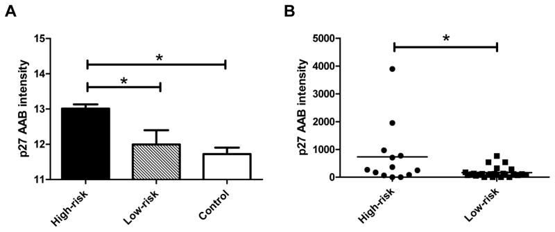
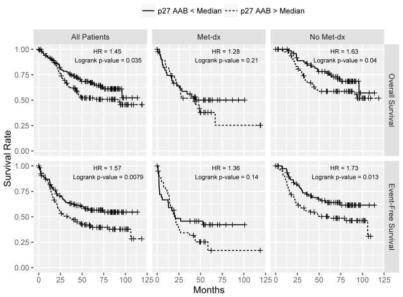
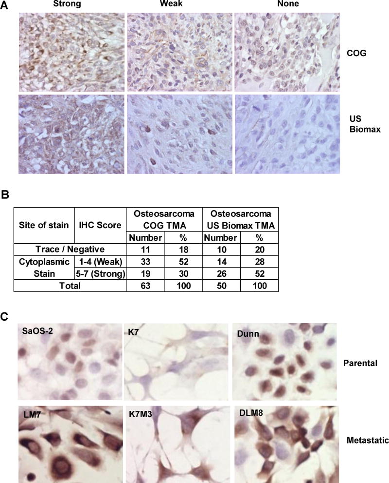
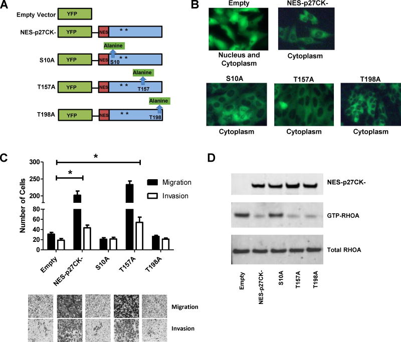
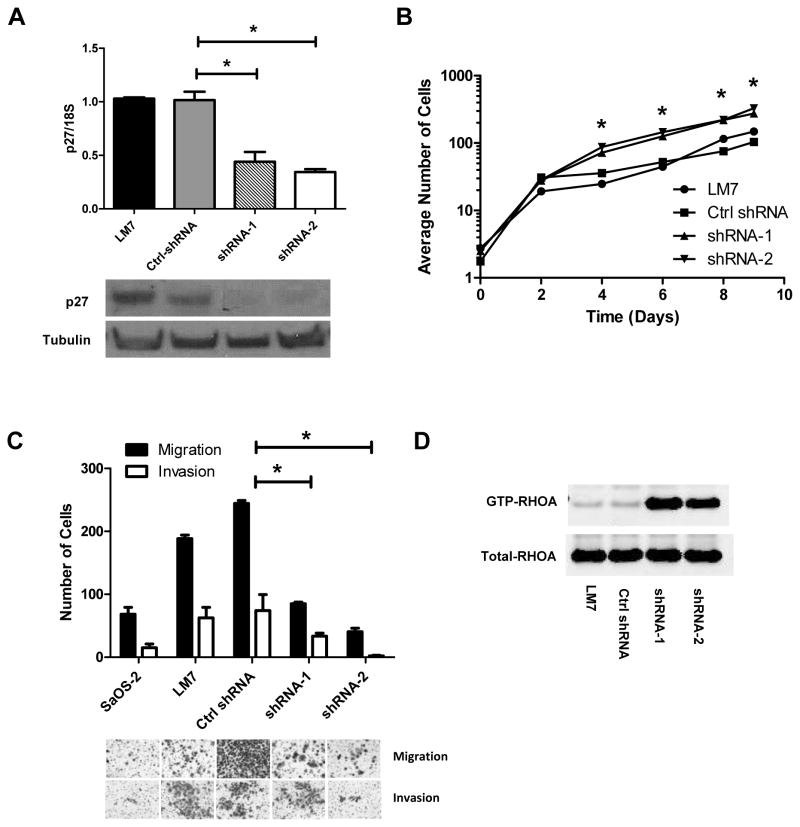
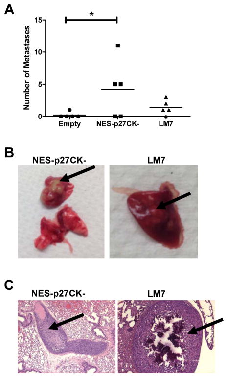
Metastatic progression is the major cause of death in osteosarcoma, the most common bone malignancy in children and young adults. However, prognostic biomarkers and efficacious targeted treatments for metastatic disease remain lacking. Using an immunoproteomic approach, we discovered that autoantibodies against the cell-cycle kinase inhibitor p27 (KIP1, CDKN1B) were elevated in plasma of high-risk osteosarcoma patients. Using a large cohort of serum samples from osteosarcoma patients (n = 233), we validated that a higher level of the p27 autoantibody significantly correlated with poor overall and event-free survival (P < 0.05). Immunohistochemical analysis also showed that p27 was mislocalized to the cytoplasm in the majority of osteosarcoma cases and in highly metastatic osteosarcoma cell lines. We demonstrated that ectopic expression of cytoplasmic p27 promoted migration and invasion of osteosarcoma cells, whereas shRNA-mediated gene silencing suppressed these effects. In addition, mutations at the p27 phosphorylation sites S10 or T198, but not T157, abolished the migratory and invasive phenotypes. Furthermore, the development of pulmonary metastases increased in mice injected with cells expressing cytoplasmic p27 compared with an empty vector control. Collectively, our findings support further investigation of p27 as a potential prognostic biomarker and therapeutic target in osteosarcoma cases exhibiting aberrant p27 subcellular localization. Cancer Res; 76(13); 4002-11. ©2016 AACR.


Conflict of interest statement

Disclosure of Potential Conflicts of Interest: The authors disclosed no potential conflicts of interest.

Figures

Figure 1
Figure 1
Identification of p27 as a tumor-associated antigen in high-risk OS. A: The normalized log fluorescent intensity of the p27 autoantibody in the pooled high-risk samples was significantly higher than those in the pooled samples from low-risk and noncancerous diseases (p < 0.05) using ProtoArrays. B: The Luminex xMAP assay validated that the p27 autoantibody levels in the individual samples of the high-risk group were significantly higher those in the low-risk group (p = 0.0134). The lines in the plot denote the average intensity values of the groups.
Figure 2
Figure 2
The Kaplan-Meier curves of the Luminex xMAP assay of the p27 autoantibody in 233 OS serum samples. Using a median intensity cutoff, a higher level of the p27 autoantibody significantly correlated with the poor overall survival (top) and event-free survival (bottom) of the OS patients (left panel). The middle and the right panels show the Kaplan-Meier curves when stratified by the initial metastasis status.
Figure 3
Figure 3
The p27 protein exhibits cytoplasmic localization in OS cases and metastatic cell lines. A: 10X immunohistochemistry images of p27 staining in two OS tumor tissue microarrays (COG: Children’s Oncology Group and US BioMax). B: A table to show the number and % of nuclear and cytoplasmic staining results in the tissue microarrays. C: 40X immunocytochemistry images show that p27 was expressed higher in the cytoplasm of the three OS metastatic cell lines (LM7, K7M3 and DLM8) when compared to the parental cells (SaOS-2, K7 and Dunn).
Figure 4
Figure 4
Functional analysis of cytoplasmic expression of p27 in OS. A: The NES constructs used in this study. All the NES-p27 constructs contain mutations in CDK binding domain (CK-) as indicated by the asterisks. NES and EYFP denote Nuclear Export Sequence and Enhanced Yellow Fluorescent Protein, respectively. The mutations in S10A, T157A and T198A are highlighted green. B: The fluorescent images (40X) indicate the subcellular localization of the empty vector (EYFP), NES-p27CK- and phosphomutant proteins in the stable clones. C: The results of the transwell cell migration and invasion assays indicate that the number of migrated and invaded cells in the NES-p27CK- and T157A cells were significantly higher than those in the empty vector control (*p < 0.05), as well as the S10A and T198A mutants. Migrated and invaded cells were quantified by counting stained cells in 3 independent microscopic fields at 6 h and 24 h, respectively. The staining was done with Cell Staining Solution included in the Cell Migration and Invasion Kit. Error bars and asterisks represent standard deviations and statistical significance (p < 0.05), respectively. D: The RHOA-GTPase activity assay to indicate the effects of cytoplasmic p27 and phosphomutations on the inhibition of the RHOA activity.
Figure 5
Figure 5
Functional analysis of shRNA-mediated gene silencing of p27 in OS. A: p27 mRNA (top) and protein (bottom) expressions were measured in LM7 cells alone and with stably transfected Ctrl-shRNA (scramble sequence), p27 shRNA-1 and p27 shRNA-2. The p27 mRNA expression was normalized to 18S ribosomal RNA. B: The growth curves show that p27-shRNA silenced mutants grew faster than the parental and control cells. The plot is in a semi-log scale where X-axis represents days of incubation. Asterisks denote statistical significance when comparing the number of cells in shRNA mutants with LM7 and control cells at each time point (*p < 0.05). C: Transwell migration and invasion assays show that the numbers of migrated and invaded cells in the p27-silcenced cells were significantly lower than the control cells. Error bars and asterisks represent standard deviations and statistical significance (*p < 0.05), respectively. D: The RHOA-GTPase activity assay shows that RHOA activation in the p27-silcenced cells was higher than in the parental or control cells.
Figure 6
Figure 6
Cytoplasmic p27 promotes the development of metastasis in an experimental mouse model. A: The number of pulmonary metastases in mice injected with SaOS-2 containing NES-p27CK- was significantly increased when compared to the empty vector control (*p < 0.05, one-tailed t-test). The lines in the plot denote the average intensity values of the groups. B: Macroscopic examination of lungs derived from the mice injected with SaOS-2 cells with NES-p27CK- and LM7 cells. C: Histological examination of metastases developed in the lung sections dissected from SaOS-2 cells with NES-p27CK- (left) and LM7 cells (right) and after H&E staining. Arrows indicate the metastatic nodules.

References

    1. Chou AJ, Geller DS, Gorlick R. Therapy for osteosarcoma: where do we go from here? Paediatr Drugs. 2008;10(5):315–27. - PubMed
    1. Link MP, Eilber F. Osteosarcoma. In: Eilber F, Poplack D, editors. Principles and Practice of Pediatric Oncology. 3. Philadelphia: Lippincott-Raven Publishers; 1997. pp. 889–920.
    1. Bielack SS, Kempf-Bielack B, Delling G, Exner GU, Flege S, Helmke K, et al. Prognostic factors in high-grade osteosarcoma of the extremities or trunk: an analysis of 1,702 patients treated on neoadjuvant cooperative osteosarcoma study group protocols. J Clin Oncol. 2002;20(3):776–90. - PubMed
    1. Kager L, Zoubek A, Potschger U, Kastner U, Flege S, Kempf-Bielack B, et al. Primary metastatic osteosarcoma: presentation and outcome of patients treated on neoadjuvant Cooperative Osteosarcoma Study Group protocols. J Clin Oncol. 2003;21(10):2011–18. - PubMed
    1. Bacci G, Briccoli A, Rocca M, Ferrari S, Donati D, Longhi A, et al. Neoadjuvant chemotherapy for osteosarcoma of the extremities with metastases at presentation: recent experience at the Rizzoli Institute in 57 patients treated with cisplatin, doxorubicin, and a high dose of methotrexate and ifosfamide. Ann Oncol. 2003;14(7):1126–34. - PubMed

Publication types

MeSH terms

Substances