

Structural grey matter changes in the substantia innominata in Alzheimer's disease and dementia with Lewy bodies: a DARTEL-VBM study
- PMID: 27197956
- PMCID: PMC5434823
- DOI: 10.1002/gps.4500
Structural grey matter changes in the substantia innominata in Alzheimer's disease and dementia with Lewy bodies: a DARTEL-VBM study
Abstract
Objectives: Several cholinergic nuclei, and in particular the nucleus basalis of Meynert, are localised to the substantia innominata in the basal forebrain. These nuclei provide major cholinergic innervation to the cerebral cortex and hippocampus, and have an essential role in cognitive function. The aim of this study was to investigate volumetric grey matter (GM) changes in the substantia innominata from structural T1 images in Alzheimer's disease (AD), dementia with Lewy bodies (DLB) and healthy older participants using voxel-based morphometry.
Methods: Participants (41 DLB, 47 AD and 39 controls) underwent 3 T T1 magnetic resonance imaging and cognitive assessments. Voxel-based morphometry analysis used SPM8 with a substantia innominata brain mask to define the subspace for voxel GM analyses. Group differences, and selected behavioural and clinical correlates, were assessed.
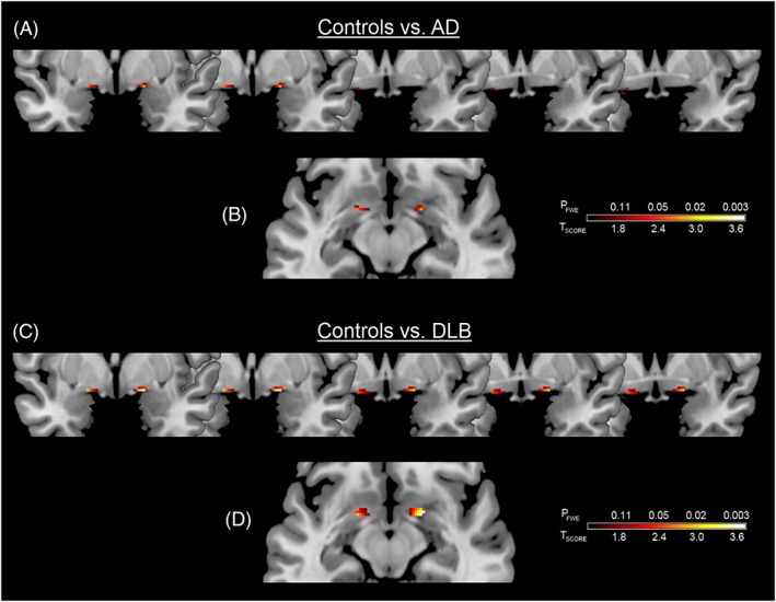
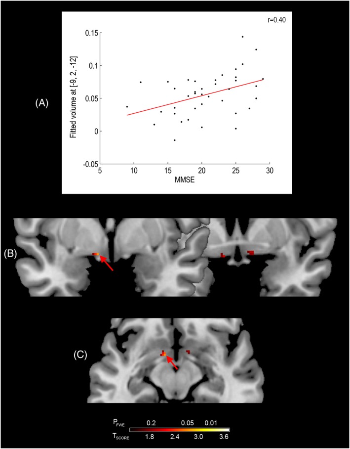
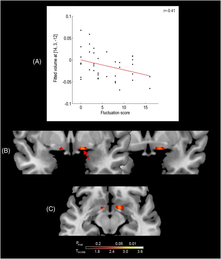
Results: Compared with that in controls, bilateral GM loss in the substantia innominata was apparent in both AD and DLB. Relative to controls, significant bilateral GM loss in the substantia innominata was observed in DLB and AD. In DLB, significant associations were also observed between substantia innominata GM volume loss, and the levels of cognitive impairment and severity of cognitive fluctuations.
Conclusions: Relative to that controls, atrophy of the substantia innominata was apparent in DLB and AD, and is associated with specific clinical manifestations in DLB. © 2016 The Authors. International Journal of Geriatric Psychiatry Published by John Wiley & Sons Ltd.
Keywords: Alzheimer's disease; DARTEL-VBM; dementia with Lewy bodies; magnetic resonance imaging; substantia innominata.
© 2016 The Authors. International Journal of Geriatric Psychiatry Published by John Wiley & Sons Ltd.
Figures

References
-
- Aarsland D, Mosimann UP, McKeith IG. 2004. Role of cholinesterase inhibitors in Parkinson's disease and dementia with Lewy bodies. J Geriatr Psychiatry Neurol 17: 164–171. - PubMed
-
- Ashburner J. 2007. A fast diffeomorphic image registration algorithm. Neuroimage 38: 95–113. - PubMed
-
- Ashburner J, Friston KJ. 2005. Unified segmentation. Neuroimage 26: 839–851. - PubMed
-
- Baxter MG, Chiba AA. 1999. Cognitive functions of the basal forebrain. Curr Opin Neurobiol 9: 178–183. - PubMed
-
- Choi SH, Jung TM, Lee JE, et al. 2012. Volumetric analysis of the substantia innominata in patients with Parkinson's disease according to cognitive status. Neurobiol Aging 33: 1265–1272. - PubMed
MeSH terms
Grants and funding
LinkOut - more resources
Full Text Sources
Other Literature Sources
Medical
