. 2016 Jun 8;19(6):891-900.
doi: 10.1016/j.chom.2016.05.004. Epub 2016 May 16.

An Infectious cDNA Clone of Zika Virus to Study Viral Virulence, Mosquito Transmission, and Antiviral Inhibitors

Affiliations

An Infectious cDNA Clone of Zika Virus to Study Viral Virulence, Mosquito Transmission, and Antiviral Inhibitors

Chao Shan et al. Cell Host Microbe. .

Abstract

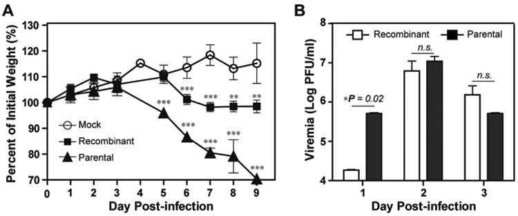
The Asian lineage of Zika virus (ZIKV) has recently caused epidemics and severe disease. Unraveling the mechanisms causing increased viral transmissibility and disease severity requires experimental systems. We report an infectious cDNA clone of ZIKV that was generated using a clinical isolate of the Asian lineage. The cDNA clone-derived RNA is infectious in cells, generating recombinant ZIKV. The recombinant virus is virulent in established ZIKV mouse models, leading to neurological signs relevant to human disease. Additionally, recombinant ZIKV is infectious for Aedes aegypti and thus provides a means to examine virus transmission. The infectious cDNA clone was further used to generate a luciferase ZIKV that exhibited sensitivity to a panflavivirus inhibitor, highlighting its potential utility for antiviral screening. This ZIKV reverse genetic system, together with mouse and mosquito infection models, may help identify viral determinants of human virulence and mosquito transmission as well as inform vaccine and therapeutic strategies.

Keywords: Zika virus; antiviral drug discovery; flavivirus; genetic system; mosquito transmission; viral virulence.


Figures

Figure. 1
Figure. 1
Construction of the infectious cDNA clone of ZIKV. (A) The strategy for constructing the full-length cDNA clone of ZIKV. Genome organization, unique restriction sites, and their nucleotide positions are shown. Five cDNA fragments from A to E (represented by thick lines) were synthesized from genomic RNA using RT-PCR to cover the complete ZIKV genome. Individual fragments were assembled to form the full-length cDNA clone of ZIKV (pFLZIKV). The complete ZIKV cDNA is positioned under the control of T7 promoter elements for in vitro transcription. An HDVr ribozyme sequence was engineered at the 3′ end of viral genome to generate an authentic 3′ end of viral RNA sequence. The numbers are the nucleotide positions based on the sequence of ZIKV strain FSS13025 (GenBank number KU955593.1). (B) Analysis of RNA transcript from pFLZIKV on a native agarose gel. A 0.8% agarose gel electrophoresis was used to analyze ZIKV RNA transcript along with a genome-length DENV-2 RNA. (C) IFA of viral protein expression in cells transfected with full-length ZIKV RNA. Vero cells were electroporated with 10 μg of genome-length ZIKV RNA. From day 3 to 6 p.t., IFA was performed to examine viral E protein expression using a mouse mAb (4G2). Green and blue represent E protein and nuclei (stained with DAPI), respectively. (D) RT-PCR analysis of progeny viral RNA. Viral RNA was extracted from culture supernatant on day 6 p.t. and used as a template for RT-PCR using ZIKV-specific primer pair 1303-F and 2552-ClaI-R (Table S2). As a negative control, a genome-length RNA containing an NS5 polymerase active site mutation (GDD mutated to AAA) was included. (E) Yield of infectious ZIKV after transfection. Viral titers from culture supernatants at indicated time points were determined by plaque assay. (F) Cytopathic effect (CPE) on Vero cells on day 6 post-transfection. CPE of Vero cells infected with parental virus on day 3 p.i. is included as a positive control.
Figure 2
Figure 2
Characterization of parental and recombinant ZIKVs in cell culture. (A) Plaque morphology of parental and recombinant ZIKVs. (B & C) Comparison of growth kinetics in Vero and C6/36 cells, respectively. Vero and C3/36 cells were infected with parental and recombinant virus at an MOI of 0.01. Viral titers were measured at indicated time points using plaque assays on Vero cells. Means and standard deviations from three independent replicates are shown. Statistics were performed using unpaired student's t-test. *significant (p value < 0.05); **highly significant (p value < 0.01). L.O.D., limitation of detection (100 PFU/ml). (D) An engineered genetic marker in the recombinant ZIKV. An SphI cleavage site, located in the viral E gene of parental virus, was knocked out in the cDNA clone to serve as a genetic marker to distinguish between recombinant virus and parental virus. A 1250-bp fragment (from nucleotides 1,303 to 2,552) spanning the SphI site was amplified using RT-PCR from RNA extracted from either recombinant virus or parental virus. The RT-PCR fragments were subjected to SphI digestion. The 1250-bp fragment derived from recombinant virus should not be cleavable by SphI; whereas the RT-PCR fragment amplified from parental viral RNA should be cleavable by SphI. The expected sizes of the digestion products are indicated. (E) Agarose gel analysis of SphI digestion products. Expected digestion pattern was observed as depicted in panel (D) was observed. The lengths of DNA fragments are indicated on the right side of the agarose gel.
Figure 3
Figure 3
Comparison of virulence in A129 mice between recombinant and parental viruses. Four-week-old A129 mice were infected with 1 × 10⁵ PFU per individual via the intraperitoneal route. Mock or infected mice (n=5 per group) were monitored for weight loss (A). The viremia at the first three days p.i. was quantified using plaque assay (B). Means and standard deviations are shown. One-way ANOVA test was performed to evaluate the statistical significance of weight differences among the mock-infected, parental virus-infected, and recombinant virus-infected mice. The unpaired student's t-test was performed to estimate the viremia differences in mice infected with recombinant and parental viruses. *, significant (p value < 0.05); **, very significant (p value < 0.01); ***, extremely significant (p value < 0.001).
Figure 4
Figure 4
Virulence of parental and recombinant ZIKVs in AG129 mice. Six week-old AG129 mice were inoculated by intraperitoneal injection with 1×10⁵ PFU (n=4 for parental virus; n=5 for recombinant virus), 1×10⁴ PFU (n=8 for parental virus; n=5 for recombinant virus), or 1×10³ PFU (n=4 for parental virus; n=5 for recombinant virus) of ZIKV. The infected mice were monitored for weight loss. Mice were euthanized once weight loss exceeded >20%. For each infection dose, weight loss and survival curves are presented. Parental and recombinant viruses and their infection doses are indicated. Values are mean percent weight compared to initial weight.
Figure 5
Figure 5
Construction of reporter ZIKV for antiviral testing. (A) Construction of a Renilla luciferase (Rluc) reporter ZIKV. The luciferase gene was in-frame fused downstream of the first 25 amino acids of the vial capsid gene (indicated as “C25”); maintaining these 25 amino acids of capsid is essential for flavivirus genome cyclization required for viral replication. The 2A peptide from foot-and-mouth disease virus (indicated as “2A”) was engineered between the C-terminus of luciferase gene and the complete open reading frame of ZIKV; the 2A sequence enables cleavage between the C-terminus of luciferase and the N-terminus of the downstream capsid protein. Silent mutations within the flavivirus-cyclization sequence (located in the amino acids 14-17 of the complete capsid gene) were engineered; the mutated nucleotides are indicated in red. (B) Plaque morphology of the recombinant ZIKV and immunostaining foci of the Rluc ZIKV. Plaque assay was performed for the wild-type ZIKV on day 4 post-infection. The Rluc ZIKV did not generate clear plaques (data not shown), but could be detected by immunostaining on day 6 post-infection. (C) Antiviral test using the reporter ZIKV. Vero cells were infected with the reporter ZIKV and treated with a pan-flavivirus nucleoside inhibitor NITD008 (Yin et al., 2009). At 48 h p.i., the cells were measured for luciferase activities. An estimated EC50 value of 2.5 μM is indicated for NITD008. (D) Anti-ZIKV activity of NITD008 using viral titer reduction assay. Vero cells were infected with wild-type ZIKV at an MOI of 0.05 in the presence of various concentrations of NITD008. At 48 h p.i., viral titers in culture fluids were quantified using plaque assay. The viral titer reduction results suggests an EC50 value of 3.6 μM.

References

    1. Alkan C, Zapata S, Bichaud L, Moureau G, Lemey P, Firth AE, Gritsun TS, Gould EA, de Lamballerie X, Depaquit J, et al. Ecuador Paraiso Escondido Virus, a New Flavivirus Isolated from New World Sand Flies in Ecuador, Is the First Representative of a Novel Clade in the Genus Flavivirus. J Virol. 2015;89:11773–11785. - PMC - PubMed
    1. Calvet G, Aguiar RS, Melo AS, Sampaio SA, de Filippis I, Fabri A, Araujo ES, de Sequeira PC, de Mendonca MC, de Oliveira L, et al. Detection and sequencing of Zika virus from amniotic fluid of fetuses with microcephaly in Brazil: a case study. The Lancet Infectious diseases 2016 - PubMed
    1. Chouin-Carneiro T, Vega-Rua A, Vazeille M, Yebakima A, Girod R, Goindin D, Dupont-Rouzeyrol M, Lourenco-de-Oliveira R, Failloux AB. Differential Susceptibilities of Aedes aegypti and Aedes albopictus from the Americas to Zika Virus. PLoS Negl Trop Dis. 2016;10:e0004543. - PMC - PubMed
    1. Dick GW, Kitchen SF, Haddow AJ. Zika virus. I. Isolations and serological specificity. Transactions of the Royal Society of Tropical Medicine and Hygiene. 1952;46:509–520. - PubMed
    1. Dudley DM, Aliota MT, Mohr EL, Weiler AM, Lehrer-Brey G, Weisgrau KL, Mohns MS, Breitbach ME, Rasheed MN, Newman CM, et al. Natural history of Asian lineage Zika virus infection in macaques. bioRxiv 2016 - PMC - PubMed

MeSH terms