. 2016 May 20:6:26291.
doi: 10.1038/srep26291.

Nrf2/Keap1 system regulates vascular smooth muscle cell apoptosis for vascular homeostasis: role in neointimal formation after vascular injury

Affiliations

Nrf2/Keap1 system regulates vascular smooth muscle cell apoptosis for vascular homeostasis: role in neointimal formation after vascular injury

Takashi Ashino et al. Sci Rep. .

Abstract

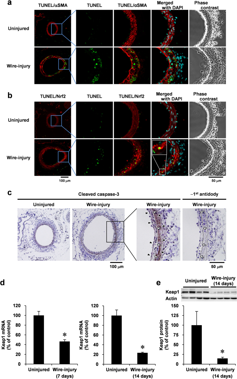
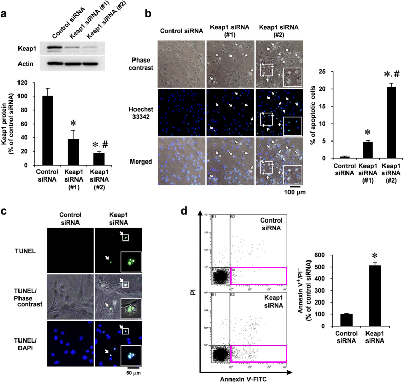
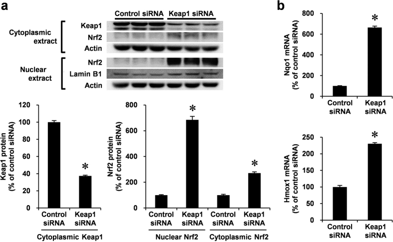
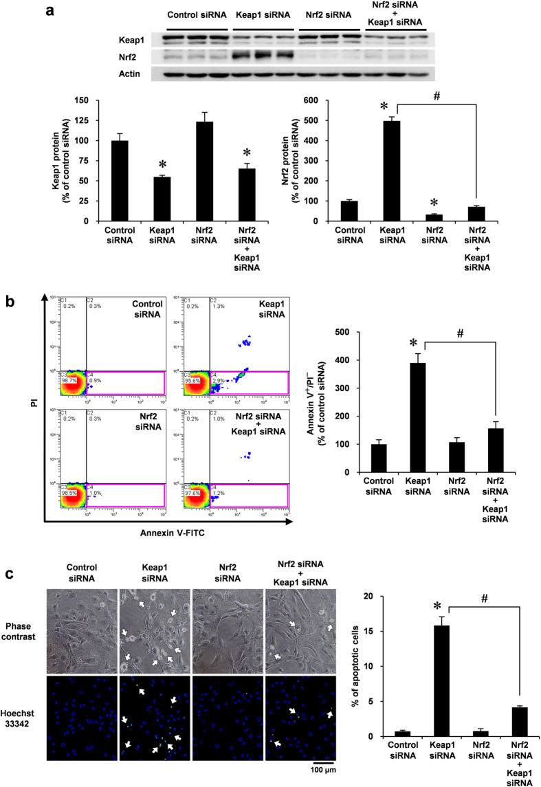
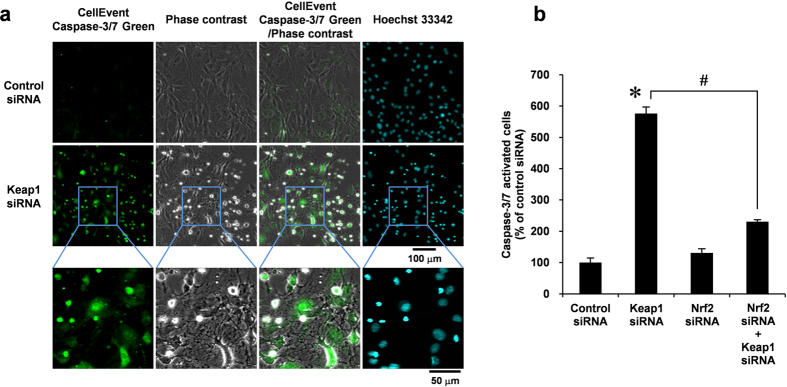
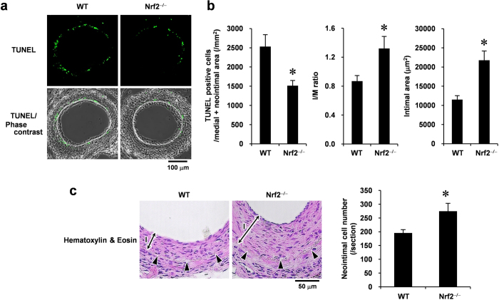
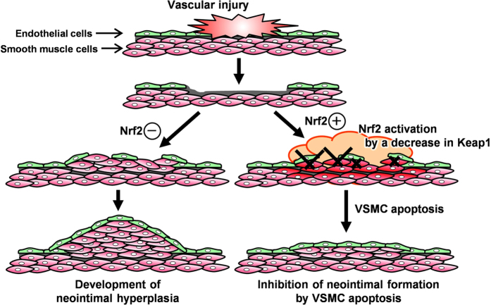
Abnormal increases in vascular smooth muscle cells (VSMCs) in the intimal region after a vascular injury is a key event in developing neointimal hyperplasia. To maintain vascular function, proliferation and apoptosis of VSMCs is tightly controlled during vascular remodeling. NF-E2-related factor 2 (Nrf2)/Kelch-like ECH-associated protein 1 (Keap1) system, a key component of the oxidative stress response that acts in maintaining homeostasis, plays an important role in neointimal hyperplasia after a vascular injury; however, the role of Nrf2/Keap1 in VSMC apoptosis has not been clarified. Here we report that 14 days after arterial injury in mice, TUNEL-positive VSMCs are detected in both the neointimal and medial layers. These layers contain cells expressing high levels of Nrf2 but low Keap1 expression. In VSMCs, Keap1 depletion induces features of apoptosis, such as positive TUNEL staining and annexin V binding. These changes are associated with an increased expression of nuclear Nrf2. Simultaneous Nrf2 depletion inhibits Keap1 depletion-induced apoptosis. At 14 days after the vascular injury, Nrf2-deficient mice demonstrated fewer TUNEL-positive cells and increased neointimal formation in the neointimal and medial areas. The results suggest that the Nrf2/Keap1 system regulates VSMC apoptosis during neointimal formation, thereby inhibiting neointimal hyperplasia after a vascular injury.


Figures

Figure 1
Figure 1. VSMC apoptosis in the middle stages of neointimal expansion after the vascular injury is associated with a high expression of Nrf2, decrease in Keap1, and caspase-3 activation.
Femoral arteries uninjured or injured (14 days later) co-stained with TUNEL (green), DAPI (blue), and anti-αSMA (red; (a)) or anti-Nrf2 (red; (b)) antibodies for immunofluorescence analysis, or stained with anti-cleaved caspase-3 antibody (c) for immunohistochemical analysis. Fluorescence images were taken by confocal microscopy under fixed exposure conditions. Arrowheads indicate cleaved caspase-3; white arrows indicate internal elastic lamina. Keap1 mRNA (d) and protein (e) levels of uninjured or injured (7 or 14 days later) femoral artery were analyzed using real-time PCR and Western blotting, respectively. Data are expressed as mean ± SEM of four vessels. *P < 0.05 vs. uninjured vessels.
Figure 2
Figure 2. Effect of Keap1 depletion in VSMC apoptosis.
(a–d) RASMCs were transiently transfected with Keap1 (#1 or #2) or control siRNA for 48 h. (a) Keap1 protein levels were analyzed by Western blotting. (b) Growth-arrested RASMCs were stained with Hoechst 33342 (blue). Apoptotic RASMCs exhibited shrunken morphology and fragmented nuclei with bright nuclear fluorescence indicated by arrows. The percentage of apoptotic cells were determined by counting shrunken cells in three separate view fields. (c) Growth-arrested RASMCs were co-stained with TUNEL (green) and DAPI (blue). Arrows indicate TUNEL positive cells. Results are representative of three independent replicates of immunofluorescence images. (d) Growth-arrested RASMCs were co-stained with annexin V–FITC and PI, and analyzed by flow cytometry. Apoptotic cells were defined by annexin V positive and PI negative cells (red square at right lower quadrants). Data are expressed as mean ± SEM of three independent experiments. *P < 0.05 vs. control siRNA. #P < 0.05 vs. Keap1 siRNA (#1).
Figure 3
Figure 3. Endogenous Keap1 knockdown by siRNA and expression of Nrf2 and its target genes, including Nqo1 and Hmox1 in VSMCs.
(a,b) RASMCs were transiently transfected with Keap1 or control siRNA for 48 h. (a) Nuclear and cytoplasmic protein levels were analyzed by Western blotting. Cytoplasmic and nuclear proteins were semi-quantified by normalizing with actin and lamin B protein, respectively. (b) mRNA levels were analyzed by real-time PCR. Data are expressed as mean ± SEM of three independent experiments. *P < 0.05 vs. control siRNA.
Figure 4
Figure 4. Effect of Nrf2 siRNA co-transfected with Keap1 siRNA in VSMC apoptosis.
(a–c) RASMCs were transiently transfected with Nrf2 or control siRNA. After 24 h, the RASMCs were additively transfected with Keap1 or control siRNA for 48 h. (a) Protein levels in the whole cell lysate were analyzed by Western blotting. (b) Growth-arrested RASMCs were co-stained with annexin V–FITC and PI and were analyzed by flow cytometry. Apoptotic cells were defined as described for Fig. 2d. (c) Growth-arrested RASMCs were stained with Hoechst 33342 (blue). The percentage of apoptotic cells were determined as described for Fig. 2b. Arrows indicate apoptotic cells. Data are expressed as mean ± SEM of three independent experiments. *P < 0.05 vs. control siRNA. #P < 0.05 vs. Keap1 siRNA alone.
Figure 5
Figure 5. Effect of Nrf2 siRNA co-transfected with Keap1 siRNA in caspase-3/7 activation in VSMCs.
(a) RASMCs were transiently transfected with Keap1 or control siRNA for 48 h. Growth-arrested RASMCs were stained with CellEvent Caspase-3/7 Green reagent (green) for 90 min. Hoechst 33342 (blue) were added 30 min before the end of the treatment. Fluorescence images were taken by confocal microscopy under fixed exposure conditions. Results are representative of three independent replicates of immunofluorescence images. (b) RASMCs were transiently transfected with Nrf2 or control siRNA. After 24 h, the RASMCs were additively transfected with Keap1 or control siRNA for 48 h. Growth-arrested RASMCs were stained with CellEvent Caspase-3/7 Green reagent, and analyzed by flow cytometry. Data are expressed as mean ± SEM of three independent experiments. *P < 0.05 vs. control siRNA. #P < 0.05 vs. Keap1 siRNA alone.
Figure 6
Figure 6. Loss of Nrf2 prevents vascular cell apoptosis and promotes neointimal formation after the vascular injury.
(a) TUNEL staining and its phase-contrast images of femoral arteries obtained from WT and Nrf2−/− mice at 14 days after injury. (b) Quantitative morphometric analysis of TUNEL-positive cells and vessel remodeling in WT and Nrf2−/− mice. I/M ratio indicates intimal area to medial area ratio. Data are expressed as mean ± SEM of three different sections from each of 10 (WT) or nine (Nrf2−/−) vessels. (c) Hematoxylin & Eosin staining of femoral arteries obtained from the WT and Nrf2−/− mice at 28 days after injury. Arrowheads indicate internal elastic lamina. I, intimal layer. The number of neointimal cells was determined by counting the nucleus. Data are expressed as mean ± SEM of sections from each of five vessels. *P < 0.05 vs. WT mice.
Figure 7
Figure 7. A proposed model for the role of Nrf2/Keap1 system in VSMCs during injury-induced neointimal expansion.
The vascular injury induces oxidative stress and a decrease in Keap1 expression, thereby activating Nrf2 in VSMCs. Stress response by Nrf2 activation promotes programmed cell death (“apoptosis”) through activation of caspase-3/7, which prevents excessively neointimal expansion for maintaining vascular function.

References

    1. Schwartz S. M. Smooth muscle migration in atherosclerosis and restenosis. J Clin Invest 100, S87–89 (1997). - PubMed
    1. Zargham R. Preventing restenosis after angioplasty: a multistage approach. Clin Sci (Lond) 114, 257–264 (2008). - PubMed
    1. Slomp J. et al. Differentiation, dedifferentiation, and apoptosis of smooth muscle cells during the development of the human ductus arteriosus. Arteriosclerosis, thrombosis, and vascular biology 17, 1003–1009 (1997). - PubMed
    1. Durand E. et al. Time courses of apoptosis and cell proliferation and their relationship to arterial remodeling and restenosis after angioplasty in an atherosclerotic rabbit model. J Am Coll Cardiol 39, 1680–1685 (2002). - PubMed
    1. Griendling K. K., Sorescu D. & Ushio-Fukai M. NAD(P)H oxidase: role in cardiovascular biology and disease. Circ Res 86, 494–501 (2000). - PubMed

Publication types

MeSH terms

Substances