Review
. 2016 Aug;25(8):1390-406.
doi: 10.1002/pro.2950. Epub 2016 Jun 8.

The ribosome in action: Tuning of translational efficiency and protein folding

Affiliations
Review

The ribosome in action: Tuning of translational efficiency and protein folding

Marina V Rodnina. Protein Sci. 2016 Aug.

Abstract

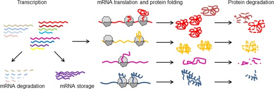
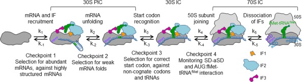
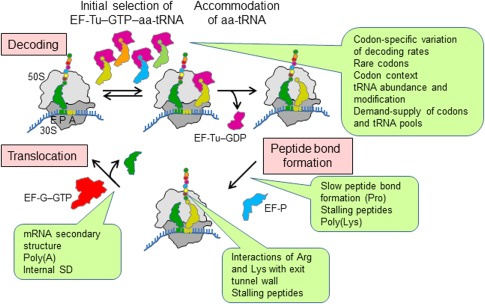
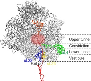
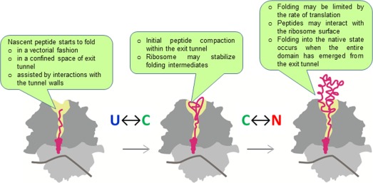
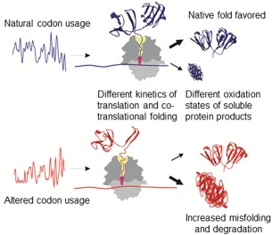
The cellular proteome is shaped by the combined activities of the gene expression and quality control machineries. While transcription plays an undoubtedly important role, in recent years also translation emerged as a key step that defines the composition and quality of the proteome and the functional activity of proteins in the cell. Among the different post-transcriptional control mechanisms, translation initiation and elongation provide multiple checkpoints that can affect translational efficiency. A multitude of specific signals in mRNAs can determine the frequency of translation initiation, choice of the open reading frame, global and local elongation velocities, and the folding of the emerging protein. In addition to specific signatures in the mRNAs, also variations in the global pools of translation components, including ribosomes, tRNAs, mRNAs, and translation factors can alter translational efficiencies. The cellular outcomes of phenomena such as mRNA codon bias are sometimes difficult to understand due to the staggering complexity of covariates that affect codon usage, translation, and protein folding. Here we summarize the experimental evidence on how the ribosome-together with the other components of the translational machinery-can alter translational efficiencies of mRNA at the initiation and elongation stages and how translation velocity affects protein folding. We seek to explain these findings in the context of mechanistic work on the ribosome. The results argue in favour of a new understanding of translation control as a hub that links mRNA homeostasis to production and quality control of proteins in the cell.

Keywords: mRNA rare codons; protein synthesis and folding; ribosome; tRNA; translational pausing.


Figures

Figure 1
Figure 1
Homeostasis of mRNA and protein. The gene‐expression pathway comprises multiple regulatory levels. Following transcription, mRNA can be degraded, stored or used for translation. Translational efficiencies of individual mRNAs (different color) depend on their sequence signatures and the pools of translational components. Protein folding frequently starts co‐translationally. When translation is completed, proteins are released from the ribosome and fulfil their functions until they are degraded by cellular proteases. Each of these steps enables the selective regulation of expression of individual mRNAs. Translational efficiency can be regulated at initiation or elongation, resulting in different pattern of ribosome distribution on the mRNA. The pace of translation can regulate protein folding (bright red and dark red protein folds).
Figure 2
Figure 2
Kinetic partitioning mechanism of mRNA selection. Checkpoint 1, initial docking complex with mRNA bound to the platform of the 30S subunit.41 Checkpoint 2, formation of the mature 30S PIC. The step indicated as mRNA unfolding may entail a number of further intermediates, for example, the formation of a stable SD‐aSD interaction, rearrangements of the 30S subunit, and possibly sampling of the initial start codon. As the latter checkpoint, mainly the secondary structure of the RBS is monitored. Recruitment of fMet‐tRNAfMet to the 30S PIC, which is not shown as a separate step, constitutes a control checkpoint for the selection of fMet‐tRNAfMet against all other aa‐tRNAs due to specific interactions with IF2. Checkpoint 3, 30S IC formation. Codon recognition is a composite step that triggers the stabilization of fMet‐tRNAfMet binding and the destabilization of IF3 binding and promotes further conformational changes in the 30S IC; checkpoint 3 selects against mismatches in the codon‐anticodon complex. Checkpoint 4, the early 70S IC. Here, the properties of the RBS are sensed.38 After GTP hydrolysis by IF2, the factors dissociate, resulting in further tightening of the 30S‐50S interactions and the formation of a mature 70S IC ready for translation elongation.40 Modified from Ref. 35.
Figure 3
Figure 3
Links between translation elongation and non‐uniform translation. Elongation entails three steps, decoding, peptide bond formation and translocation. During decoding, EF‐Tu in bacteria (or eEF1α in eukaryotes) delivers aa‐tRNA to the A site of the ribosome. These factors are GTPases that in their GTP‐bound conformation form a high‐affinity ternary complex with aa‐tRNA and GTP which, in turn, binds to the ribosome and, after GTP hydrolysis, releases aa‐tRNA to accommodate in the PTC. The ribosome selects an aa‐tRNA that is cognate to the codon in the A site (yellow) among other aa‐tRNAs. These can be almost‐cognate (orange), near‐cognate (green) or non‐cognate (blue). Peptide bond formation between aa‐tRNA in the A site and pept‐tRNA in the P site is catalyzed by the ribosome and usually does not require auxiliary proteins. The peptidyl transfer reaction between two Pro residues requires EF‐P (eIF5A in eukaryotes). Translocation is catalyzed by EF‐G (eEF2 in eukaryotes) at the cost of GTP hydrolysis. Ribosomes, aa‐tRNA and factors are all active players, and the exact interplay between them determines the actual speed and fidelity of translation. Callouts summarize potential sources of translational non‐uniformity at each step of the elongation cycle.
Figure 4
Figure 4
Schematic representation of a cross‐section of the ribosome with the nascent peptide. The peptide exit tunnel (overall length 100 Å) can be separated into three folding zones, as indicated.154 Ribosomal proteins uL4 and uL22 form the constriction. Protein uL23 and residues of 23S rRNA in the upper tunnel region (indicated by glow) can signal events in the tunnel to the PTC and the ribosome surface in the vicinity of the exit port.115
Figure 5
Figure 5
Effect of the ribosome on protein folding. U, unfolded protein; C, compact transient state or folding intermediate; N, native fold. Callouts summarize the potential effects at each step.
Figure 6
Figure 6
Synonymous codon usage directs co‐translational folding toward different protein conformations Reproduced from Ref. 156 with permission.

Similar articles

Cited by

References

    1. Ciandrini L, Stansfield I, Romano MC (2013) Ribosome traffic on mRNAs maps to gene ontology: genome‐wide quantification of translation initiation rates and polysome size regulation. PLoS Comput Biol 9:e1002866. - PMC - PubMed
    1. Siwiak M, Zielenkiewicz P (2013) Transimulation—protein biosynthesis web service. PLoS One 8:e73943. - PMC - PubMed
    1. Mitarai N, Sneppen K, Pedersen S (2008) Ribosome collisions and translation efficiency: optimization by codon usage and mRNA destabilization. J Mol Biol 382:236–245. - PubMed
    1. Subramaniam AR, Zid BM, O'Shea EK (2014) An integrated approach reveals regulatory controls on bacterial translation elongation. Cell 159:1200–1211. - PMC - PubMed
    1. Richter JD, Sonenberg N (2005) Regulation of cap‐dependent translation by eIF4E inhibitory proteins. Nature 433:477–480. - PubMed

Publication types

LinkOut - more resources