Review
. 2016 Oct 26;116(20):12730-12757.
doi: 10.1021/acs.chemrev.6b00125. Epub 2016 May 20.

Retroviral DNA Integration

Affiliations
Review

Retroviral DNA Integration

Paul Lesbats et al. Chem Rev. .

Abstract

The integration of a DNA copy of the viral RNA genome into host chromatin is the defining step of retroviral replication. This enzymatic process is catalyzed by the virus-encoded integrase protein, which is conserved among retroviruses and LTR-retrotransposons. Retroviral integration proceeds via two integrase activities: 3'-processing of the viral DNA ends, followed by the strand transfer of the processed ends into host cell chromosomal DNA. Herein we review the molecular mechanism of retroviral DNA integration, with an emphasis on reaction chemistries and architectures of the nucleoprotein complexes involved. We additionally discuss the latest advances on anti-integrase drug development for the treatment of AIDS and the utility of integrating retroviral vectors in gene therapy applications.


Conflict of interest statement

The authors declare no competing financial interest.

Figures

Figure 1
Figure 1
Retroviral DNA integration pathway: reactions catalyzed by IN (a, blue arrows) and host cell enzymes (b, red arrows). The intasome contains a multimer of IN (gray oval) assembled on vDNA ends. Following 3′-processing and nuclear entry, the cleaved intasome complex engages cellular chromosomal DNA, forming the TCC. Insertion of the 3′ vDNA ends into host DNA results in formation of the STC with hemi-integrated vDNA. Formation of the stable provirus further requires disassembly of the STC and repair of the strand discontinuities flanking vDNA by the sequential actions of a DNA polymerase, 5′-flap endonuclease, and DNA ligase. Red dots represent magnesium ions in the intasome active sites.
Figure 2
Figure 2
Domain organization of HIV-1 IN. (a) Schematic of the protein sequence with the NTD, CCD, and CTD shown as boxes (top). Structures of the individual domains determined in isolation (from left to right PDB IDs 1WJC, 1ITG, and 1IHV). Protein chains are shown as cartoons, with zinc-coordinating residues His12, His16, Cys40, and Cys43 and active site residues Asp64 and Asp116 shown as sticks with carbon atoms in red. (b) Structure of a two-domain HIV-1 IN construct (PDB ID 1K6Y). Details of the key NTD–CCD interface are shown as a blown up image to the right. Selected amino acid residues are indicated. Dashes represent hydrogen bonds; gray spheres are Zn2+ ions. All structural images in this review were prepared using PyMOL software (http://www.pymol.org).
Figure 3
Figure 3
Structure of the PFV intasome. (a) Cleaved intasome complex (PDB ID 3L2Q) shown in two orthogonal views. Inner IN chains are colored green and cyan, and outer chains are yellow. Individual IN domains are indicated; invariant IN active site carboxylates are shown as red sticks; vDNA is shown as cartoons with transferred (i.e., the strand that IN acts upon) and nontransferred strands at each end shown in magenta and beige, respectively. (b) Schematic of the intasomal tetramer with IN domains shown as ovals and colored by chain; curved lines represent NTD–CCD (cyan and green) and CCD–CTD (black) linkers. Red circles represent inner IN chain active sites. (c) View on the IN active site prior to 3′-processing. Active site carboxylates and catalytic metal ions are respectively shown as sticks and gray spheres. The invariant vDNA CA nucleotides and their complements are letter-coded; the scissile 3′ dinucleotide is indicated.
Figure 4
Figure 4
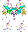
Target DNA capture by the PFV intasome. (a) Crystal structure of the PFV TCC (PDB ID 3OS1). (b) Conformation of vDNA and target DNA in the TCC (left) and STC (right, PDB ID 3OS0), with protein chains hidden for clarity.
Figure 5
Figure 5
PFV intasome engaged with a mononucleosome. (a) Pseudoatomic model assembled by rigid body docking of the PFV intasome (PDB ID 3L2Q) and nucleosome (PDB ID 1KX5) structures into the cryo-EM map of the complex (EMDB ID 2992) and shown in two orientations. Individual histones are color coded as indicated. (b) Blow up of the regions boxed in the left side of panel a showing IN contacts with H2A and H2B histones (left) and with the second gyre of the nucleosomal DNA (right).
Figure 6
Figure 6
Configurations of the IN active site leading to 3′-processing (a, PDB ID 4E7I) and strand transfer (b, PDB ID 4E7K). Direction of nucleophilic attack by a water molecule (W, panel a) or 3′-hydroxyl of vDNA (panel b) is indicated with blue arrows marked SN2. Red spheres represent water molecules; lower TCC X-ray data resolution did not allow refinement of water molecules in panel b.
Figure 7
Figure 7
Architecture of the α and β-retroviral intasomes. (a) Crystal structure of the ASLV STC (PDB ID 5EJK) in two orientations. Target DNA is hidden in the top panel. The core IN tetramer containing inner and outer IN subunits and a pair of synaptic CTDs from the flanking IN chains is indicated. (b) Schematic of the intasomal IN octamer with IN domains shown as ovals and colored by chain. For clarity, CTDs and NTDs belonging to the outer IN subunits and NTDs from the flanking subunits present in the structures are not indicated in panel a or shown in panel c. (c) Pseudo-atomic model of MMTV intasome based on cryo-EM data (PDB ID 3JCA and EMDB ID 6441).
Figure 8
Figure 8
Interaction of LEDGF/p75 and ALLINIs with HIV-1 IN. (a) Schematic of LEDGF/p75 and p52 organization with NLS, AT-hooks, and structural domains (PWWP and IBD) shown as boxes. (b) Crystal structure of the HIV-1 IN CCD dimer in complex with LEDGF/p75 IBD (PDB ID 2B4J). (c) Details of the protein–protein interactions (rotated ∼90° counterclockwise from the boxed region in panel b). (d) Chemical structures of selected ALLINIs; chemical groups mimicking LEDGF/p75 Asp366 and Ile365 are shown in red and blue, respectively. (e) Crystal structure of ALLINI BI-D in complex with HIV-1 IN CCD (PDB ID 4ID1), aligned with the panel c projection.
Figure 9
Figure 9
Interaction of BET proteins with MLV IN. (a) Schematic of domain compositions of BRD2, BRD3, and BRD4. (b) Solution structure of BRD4 ET domain in complex with the EBM of MLV IN (PDB ID 2N3K) shown in two orientations. BRD4 and IN residues participating in the hydrophobic core of the interface are shown as sticks and are indicated.
Figure 10
Figure 10
Inhibitors of HIV-1 IN strand transfer activity. (a) Chemical structures of a diketo acid (L-731,988) and the clinical INSTIs RAL, EVG, and DTG. Metal chelating atoms and halobenzyl moieties of the INSTIs are shown in red and blue, respectively. (b) Active site of the PFV intasome prior to (top) and after binding RAL (middle) or DTG (bottom). The 310 helix that participates in the interactions with the INSTIs is indicated as η.

References

    1. Rous P. A Sarcoma of the Fowl Transmissible by an Agent Separable from the Tumor Cells. J. Exp. Med. 1911, 13 (4), 397–411. 10.1084/jem.13.4.397. - DOI - PMC - PubMed
    1. Ellermann V.; Bang O. Experimentelle Leukämie Bei Hühnern. Zentralbl. Bakteriol. Parasitenkd. Infectionskr. Hyg. Abt. Orig. 1908, 46, 595–609.
    1. Poiesz B. J.; Ruscetti F. W.; Gazdar A. F.; Bunn P. A.; Minna J. D.; Gallo R. C. Detection and Isolation of Type C Retrovirus Particles from Fresh and Cultured Lymphocytes of a Patient with Cutaneous T-Cell Lymphoma. Proc. Natl. Acad. Sci. U. S. A. 1980, 77 (12), 7415–7419. 10.1073/pnas.77.12.7415. - DOI - PMC - PubMed
    1. Barre-Sinoussi F.; Chermann J. C.; Rey F.; Nugeyre M. T.; Chamaret S.; Gruest J.; Dauguet C.; Axler-Blin C.; Vezinet-Brun F.; Rouzioux C.; et al. Isolation of a T-Lymphotropic Retrovirus from a Patient at Risk for Acquired Immune Deficiency Syndrome (AIDS). Science 1983, 220 (4599), 868–871. 10.1126/science.6189183. - DOI - PubMed
    1. Popovic M.; Sarngadharan M. G.; Read E.; Gallo R. C. Detection, Isolation, and Continuous Production of Cytopathic Retroviruses (HTLV-III) from Patients with AIDS and Pre-AIDS. Science 1984, 224 (4648), 497–500. 10.1126/science.6200935. - DOI - PubMed

Publication types