

The Effects of Tumstatin on Vascularity, Airway Inflammation and Lung Function in an Experimental Sheep Model of Chronic Asthma
- PMID: 27199164
- PMCID: PMC4873797
- DOI: 10.1038/srep26309
The Effects of Tumstatin on Vascularity, Airway Inflammation and Lung Function in an Experimental Sheep Model of Chronic Asthma
Abstract
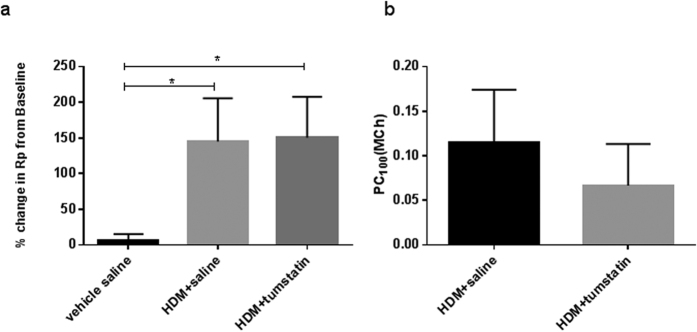
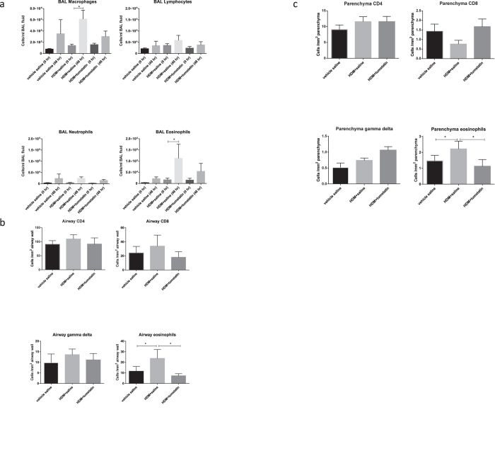
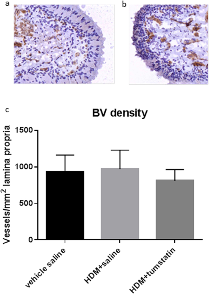
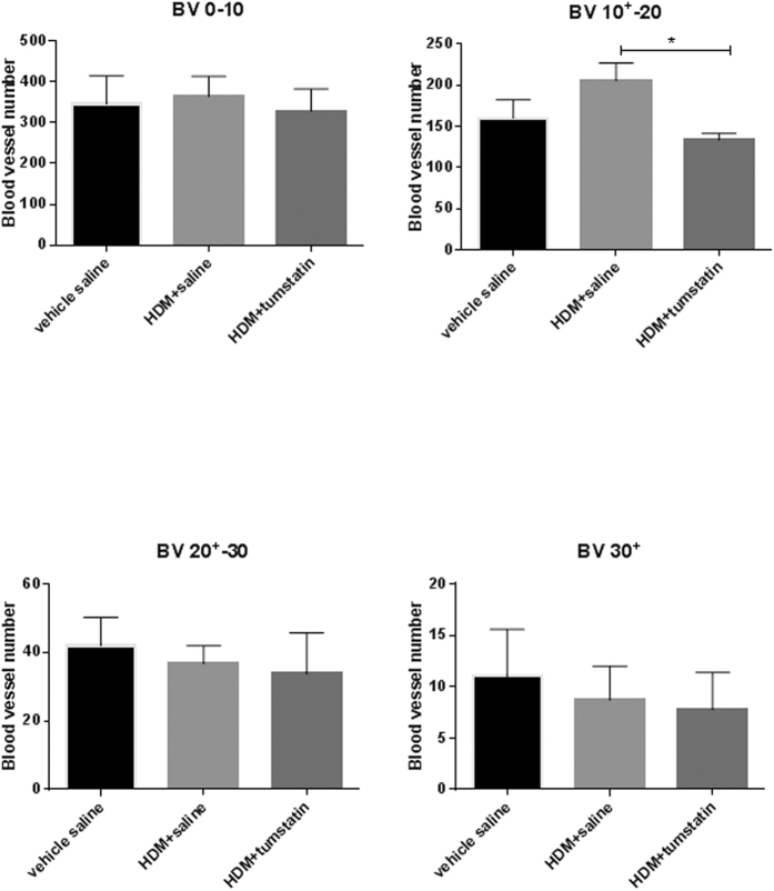
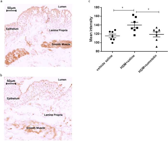
Tumstatin, a protein fragment of the alpha-3 chain of Collagen IV, is known to be significantly reduced in the airways of asthmatics. Further, there is evidence that suggests a link between the relatively low level of tumstatin and the induction of angiogenesis and inflammation in allergic airway disease. Here, we show that the intra-segmental administration of tumstatin can impede the development of vascular remodelling and allergic inflammatory responses that are induced in a segmental challenge model of experimental asthma in sheep. In particular, the administration of tumstatin to lung segments chronically exposed to house dust mite (HDM) resulted in a significant reduction of airway small blood vessels in the diameter range 10(+)-20 μm compared to controls. In tumstatin treated lung segments after HDM challenge, the number of eosinophils was significantly reduced in parenchymal and airway wall tissues, as well as in the bronchoalveolar lavage fluid. The expression of VEGF in airway smooth muscle was also significantly reduced in tumstatin-treated segments compared to control saline-treated segments. Allergic lung function responses were not attenuated by tumstatin administration in this model. The data are consistent with the concept that tumstatin can act to suppress vascular remodelling and inflammation in allergic airway disease.
Figures
Similar articles
-
Tumstatin regulates the angiogenic and inflammatory potential of airway smooth muscle extracellular matrix.J Cell Mol Med. 2017 Dec;21(12):3288-3297. doi: 10.1111/jcmm.13232. Epub 2017 Jun 13. J Cell Mol Med. 2017. PMID: 28608951 Free PMC article.
-
Reduction of tumstatin in asthmatic airways contributes to angiogenesis, inflammation, and hyperresponsiveness.Am J Respir Crit Care Med. 2010 Jan 15;181(2):106-15. doi: 10.1164/rccm.200904-0631OC. Epub 2009 Oct 29. Am J Respir Crit Care Med. 2010. PMID: 19875687
-
Airway remodelling and inflammation in sheep lungs after chronic airway challenge with house dust mite.Clin Exp Allergy. 2005 Feb;35(2):146-52. doi: 10.1111/j.1365-2222.2005.02137.x. Clin Exp Allergy. 2005. PMID: 15725184
-
The structural and functional consequences of chronic allergic inflammation of the airways.Ciba Found Symp. 1997;206:71-86; discussion 86-9, 106-10. doi: 10.1002/9780470515334.ch5. Ciba Found Symp. 1997. PMID: 9257006 Review.
-
Angioplasticity in asthma.Biochem Soc Trans. 2009 Aug;37(Pt 4):805-10. doi: 10.1042/BST0370805. Biochem Soc Trans. 2009. PMID: 19614598 Free PMC article. Review.
Cited by
-
Role of animal models in biomedical research: a review.Lab Anim Res. 2022 Jul 1;38(1):18. doi: 10.1186/s42826-022-00128-1. Lab Anim Res. 2022. PMID: 35778730 Free PMC article. Review.
-
Pathobiology of Airway Remodeling in Asthma: The Emerging Role of Integrins.J Asthma Allergy. 2022 May 11;15:595-610. doi: 10.2147/JAA.S267222. eCollection 2022. J Asthma Allergy. 2022. PMID: 35592385 Free PMC article. Review.
-
Basement membranes in obstructive pulmonary diseases.Matrix Biol Plus. 2021 Nov 12;12:100092. doi: 10.1016/j.mbplus.2021.100092. eCollection 2021 Dec. Matrix Biol Plus. 2021. PMID: 34877523 Free PMC article. Review.
-
Genetic and Environmental Contributions to Serological Biomarkers of Extracellular Matrix Remodeling in Asthma: A Twin Study.Clin Transl Allergy. 2025 Aug;15(8):e70089. doi: 10.1002/clt2.70089. Clin Transl Allergy. 2025. PMID: 40760731 Free PMC article.
-
Tumstatin regulates the angiogenic and inflammatory potential of airway smooth muscle extracellular matrix.J Cell Mol Med. 2017 Dec;21(12):3288-3297. doi: 10.1111/jcmm.13232. Epub 2017 Jun 13. J Cell Mol Med. 2017. PMID: 28608951 Free PMC article.
References
-
- Chetta A., Zanini A., Torre O. & Olivieri D. Vascular remodelling and angiogenesis in asthma: Morphological aspects and pharmacological modulation. Inflamm. Allergy Drug Targets 6, 41–45 (2007). - PubMed
-
- Hashimoto M., Tanaka H. & Abe S. Quantitative analysis of bronchial wall vascularity in the medium and small airways of patients with asthma and COPD. Chest 127, 965–972 (2005). - PubMed
-
- Li X. & Wilson J. W. Increased vascularity of the bronchial mucosa in mild asthma. Am. J. Respir. Crit. Care Med. 156, 229–233 (1997). - PubMed
-
- Siddiqui S., Hollins F., Saha S. & Brightling C. E. Inflammatory cell microlocalisation and airway dysfunction: cause and effect? Eur. Respir. J. 30, 1043–1056 (2007). - PubMed
-
- Wilson J. W. & Hii S. The importance of the airway microvasculature in asthma. Curr Opin Allergy Clin Immunol 6, 51–55 (2006). - PubMed
Publication types
MeSH terms
Substances
LinkOut - more resources
Full Text Sources
Other Literature Sources
Medical
