

Skeletal muscle fiber type: using insights from muscle developmental biology to dissect targets for susceptibility and resistance to muscle disease
- PMID: 27199166
- PMCID: PMC5180455
- DOI: 10.1002/wdev.230
Skeletal muscle fiber type: using insights from muscle developmental biology to dissect targets for susceptibility and resistance to muscle disease
Abstract
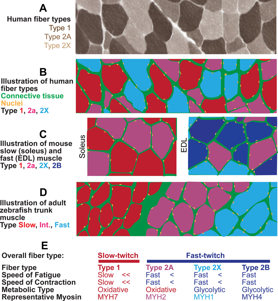
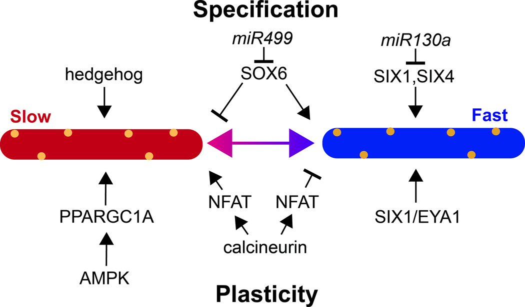
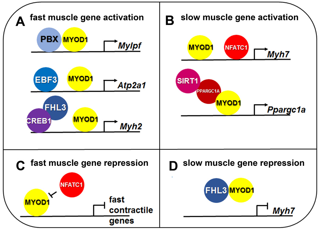
Skeletal muscle fibers are classified into fiber types, in particular, slow twitch versus fast twitch. Muscle fiber types are generally defined by the particular myosin heavy chain isoforms that they express, but many other components contribute to a fiber's physiological characteristics. Skeletal muscle fiber type can have a profound impact on muscle diseases, including certain muscular dystrophies and sarcopenia, the aging-induced loss of muscle mass and strength. These findings suggest that some muscle diseases may be treated by shifting fiber type characteristics either from slow to fast, or fast to slow phenotypes, depending on the disease. Recent studies have begun to address which components of muscle fiber types mediate their susceptibility or resistance to muscle disease. However, for many diseases it remains largely unclear why certain fiber types are affected. A substantial body of work has revealed molecular pathways that regulate muscle fiber type plasticity and early developmental muscle fiber identity. For instance, recent studies have revealed many factors that regulate muscle fiber type through modulating the activity of the muscle regulatory transcription factor MYOD1. Future studies of muscle fiber type development in animal models will continue to enhance our understanding of factors and pathways that may provide therapeutic targets to treat muscle diseases. WIREs Dev Biol 2016, 5:518-534. doi: 10.1002/wdev.230 For further resources related to this article, please visit the WIREs website.
© 2016 Wiley Periodicals, Inc.
Figures
References
-
- Schiaffino S, Reggiani C. Fiber types in mammalian skeletal muscles. Physiol Rev. 2011;91(4):1447–1531. - PubMed
-
- Bassel-Duby R, Olson EN. Signaling pathways in skeletal muscle remodeling. Annu Rev Biochem. 2006;75:19–37. - PubMed
-
- Pette D, Staron RS. Myosin isoforms, muscle fiber types, and transitions. Microsc Res Tech. 2000;50(6):500–509. - PubMed
-
- Hernandez LP, Patterson SE, Devoto SH. The development of muscle fiber type identity in zebrafish cranial muscles. Anat Embryol (Berl) 2005;209(4):323–334. - PubMed
-
- Guth L, Samaha FJ. Qualitative differences between actomyosin ATPase of slow and fast mammalian muscle. Exp Neurol. 1969;25(1):138–152. - PubMed
Publication types
MeSH terms
Substances
Grants and funding
LinkOut - more resources
Full Text Sources
Other Literature Sources
Medical
