. 2016 Jul 1;129(13):2493-9.
doi: 10.1242/jcs.189910. Epub 2016 May 19.

Meiotic onset is reliant on spatial distribution but independent of germ cell number in the mouse ovary

Affiliations

Meiotic onset is reliant on spatial distribution but independent of germ cell number in the mouse ovary

Ripla Arora et al. J Cell Sci. .

Abstract

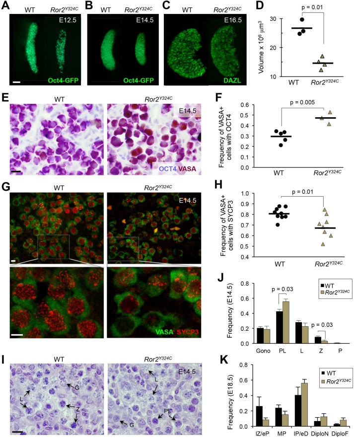
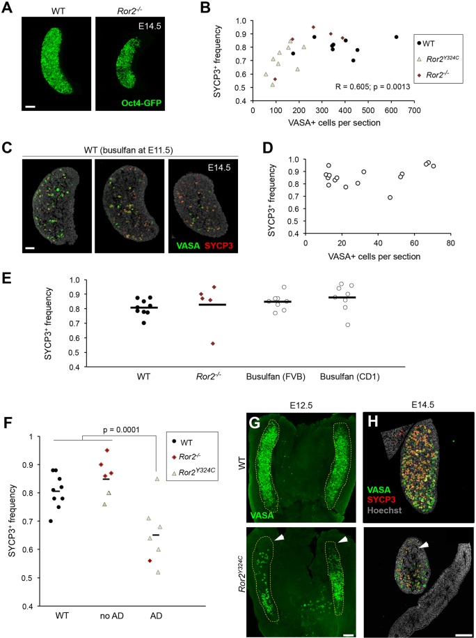
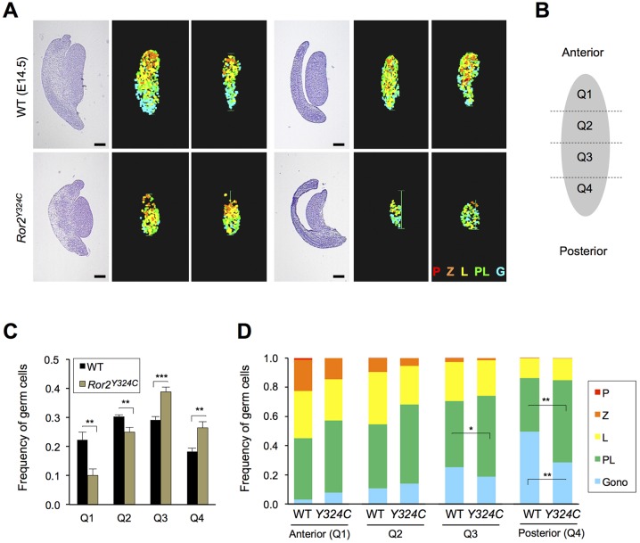
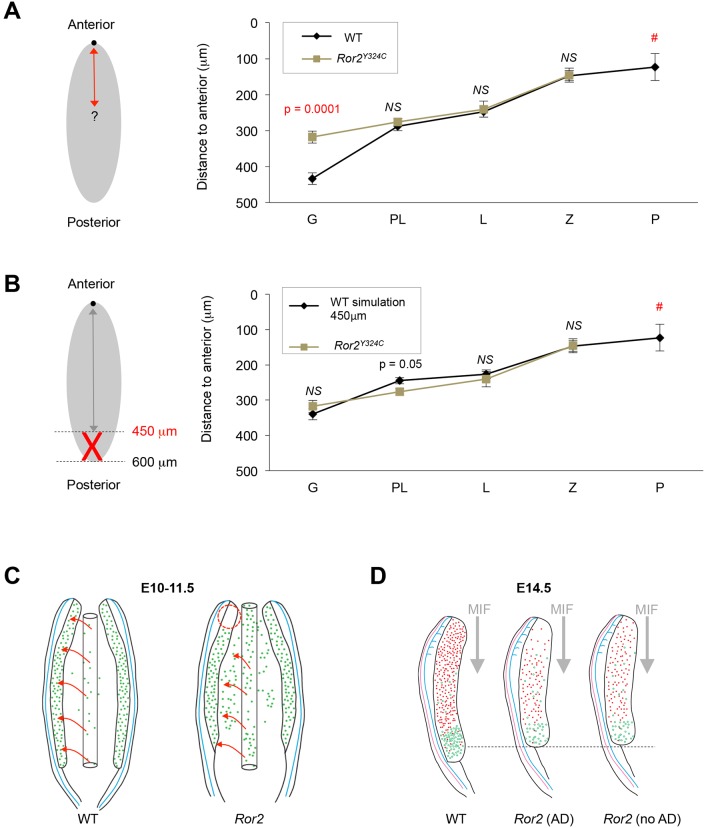
Mouse ovarian germ cells enter meiosis in a wave that propagates from anterior to posterior, but little is known about contribution of germ cells to initiation or propagation of meiosis. In a Ror2 mutant with diminished germ cell number and migration, we find that overall timing of meiotic initiation is delayed at the population level. We use chemotherapeutic depletion to exclude a profoundly reduced number of germ cells as a cause for meiotic delay. We rule out sex reversal or failure to specify somatic support cells as contributors to the meiotic phenotype. Instead, we find that anomalies in the distribution of germ cells as well as gonad shape in mutants contribute to aberrant initiation of meiosis. Our analysis supports a model of meiotic initiation via diffusible signal(s), excludes a role for germ cells in commencing the meiotic wave and furnishes the first phenotypic demonstration of the wave of meiotic entry. Finally, our studies underscore the importance of considering germ cell migration defects while studying meiosis to discern secondary effects resulting from positioning versus primary meiotic entry phenotypes.

Keywords: Germ cell; Gonad; Meiosis; Migration; Ror2; Wave.


Conflict of interest statement

Competing interests

The authors declare no competing or financial interests.

Figures

Fig. 1.
Fig. 1.
Meiotic entry is delayed in Ror2Y324C/Y324C ovaries. (A–D) Smaller ovaries and diminished germ cells in Ror2Y324C/Y324C; Oct4–GFP at E12.5 (A), E14.5 (B), and DAZL at E16.5 (C) and volume measurement (D, P=0.01, t-test). (E,F) Higher frequency of VASA+ OCT4+ germ cells in mutant ovaries (P=0.005, t-test). (G,H). Lower frequency of SYCP3+ germ cells in mutant ovaries (P=0.01, t-test). (I) H&E sections of E14.5 ovaries. Arrows indicate stages of meiotic prophase. (J,K) Quantification of I. n=6 at E14.5 (J) and n=3 at E18.5 (K). Data represented as mean±s.e.m. G/Gono, gonocytes; PL, pre-leptotene; L, leptotene; Z, zygotene; P, pachytene; lZ/eP, late-zygotene/early-pachytene; MP, mid-pachytene; IP/eD, intermediate-pachytene/early-diplotene; DiploN, naked-diplotene; DiploF, diplotene-forming follicle. Scale bars: 100 µm in A–C; 10 µm in E,G,I.
Fig. 2.
Fig. 2.
Diminished number of germ cells does not delay meiotic initiation. (A) Smaller size and reduced number of germ cells (Oct4–GFP) in whole Ror2−/− ovaries. (B) Scatter plot shows correlation (r=0.605, P=0.0013) between number of germ cells (VASA+) per section and frequency of SYCP3+ germ cells. (C) Busulfan-treated ovary sections. (D) Frequency of SYCP3+ germ cells at E14.5 did not decrease in busulfan-treated ovaries. (E) Percentage of SYCP3+ germ cells was similar between WT, Ror2−/− and busulfan treatment. (F) Data in Fig. 2B re-plotted, segregating ovaries with and without anterior defect (AD) in Ror mutants. (G) Anterior depletion of germ cells (VASA, green) in Ror2Y324C mutants. (H) Sagittal sections show germ cells (VASA, green) that have entered meiosis (SYCP3, red) at E14.5. White arrowheads, anterior defects. Images are oriented with anterior at the top. Scale bars: 100 µm in A,G,H; 50 µm in C.
Fig. 3.
Fig. 3.
Aberrant distribution of germ cells in the ovary causes meiotic entry defect. (A) Histologic sections with H&E stain at left and heat maps indicating meiotic progression on right. (B) Diagram showing division of ovaries into quadrants. (C) Relative frequency of germ cells counted in each quadrant in WT versus Ror2Y324C/Y324C, n=6 embryos. Data represented as mean±s.e.m. (D) Distribution of germ cells scored in each stage of meiotic prophase for each quadrant, n=6 WT and n=6 Ror2Y324C/Y324C ovaries. Gono, gonocytes; PL, preleptotene stage; L, leptotene stage; Z, zygotene stage; P, pachytene stage. ***P<0.001, **P<0.01, *P<0.05 by t-test. Scale bars: 200 µm.
Fig. 4.
Fig. 4.
Anterior loss of germ cells and axial compression of the gonad account for overall meiotic delay in Ror2 mutants. (A) Comparison of mean measured distance between ovary anterior and meiotic progression of germ cells in Ror2Y324C and WT (n=6 each). (B) Simulation of axial shortening of Ror2 gonads using WT dataset. Gono, gonocytes; PL, preleptotene stage; L, leptotene stage; Z, zygotene stage; P, pachytene stage. NS, not significant (t-test); #, none detected in Ror2Y324C. Data represented as mean±s.e.m. (C) Model for germ cell migration defects in Ror2 mutants that lead to the observed anterior defect. Arrows indicate path of germ cell migration from hindgut to the genital ridges. (D) Schematic for meiotic initiation in Ror2 mutants with and without anterior germ cell defects (AD). Red dots represent germ cells undergoing meiosis whereas green are pre-meiotic germ cells.

Similar articles

Cited by

References

    1. Abby E., Tourpin S., Ribeiro J., Daniel K., Messiaen S., Moison D., Guerquin J., Gaillard J-C, Armengaud J., Langa F. et al. (2016). Implementation of meiosis prophase I programme requires a conserved retinoid-independent stabilizer of meiotic transcripts. Nat. Commun. 7, 10324 10.1038/ncomms10324 - DOI - PMC - PubMed
    1. Arora R., Altman E., Tran N. D. and Laird D. J. (2014). Novel domains of expression for orphan receptor tyrosine kinase Ror2 in the human and mouse reproductive system. Dev. Dyn. 243, 1037-1045. 10.1002/dvdy.24138 - DOI - PMC - PubMed
    1. Boiani M., Kehler J. and Scholer H. R. (2004). Activity of the germline-specific Oct4-GFP transgene in normal and clone mouse embryos. Methods Mol. Biol. 254, 1-34. 10.1385/1-59259-741-6:001 - DOI - PubMed
    1. Bowles J., Knight D., Smith C., Wilhelm D., Richman J., Mamiya S., Yashiro K., Chawengsaksophak K., Wilson M. J., Rossant J. et al. (2006). Retinoid signaling determines germ cell fate in mice. Science 312, 596-600. 10.1126/science.1125691 - DOI - PubMed
    1. Buehr M., McLaren A., Bartley A. and Darling S. (1993). Proliferation and migration of primordial germ cells in We/We mouse embryos. Dev. Dyn. 198, 182-189. 10.1002/aja.1001980304 - DOI - PubMed

Publication types

Substances