. 2016 Jun 24;119(1):55-68.
doi: 10.1161/CIRCRESAHA.116.308304. Epub 2016 May 19.

CD74-Downregulation of Placental Macrophage-Trophoblastic Interactions in Preeclampsia

Affiliations

CD74-Downregulation of Placental Macrophage-Trophoblastic Interactions in Preeclampsia

Lukasz Przybyl et al. Circ Res. .

Abstract

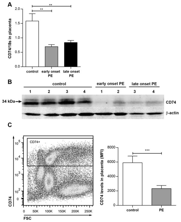
Rationale: We hypothesized that cluster of differentiation 74 (CD74) downregulation on placental macrophages, leading to altered macrophage-trophoblast interaction, is involved in preeclampsia.

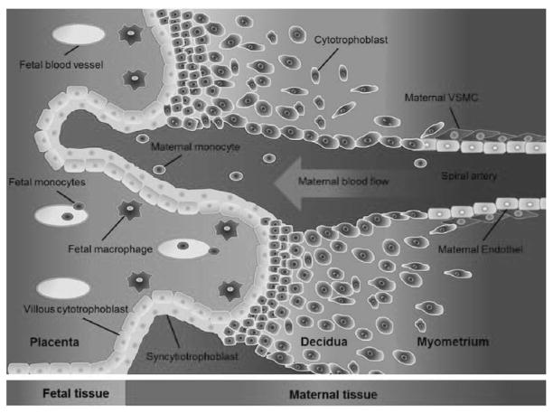
Objective: Preeclamptic pregnancies feature hypertension, proteinuria, and placental anomalies. Feto-placental macrophages regulate villous trophoblast differentiation during placental development. Disturbance of this well-balanced regulation can lead to pathological pregnancies.

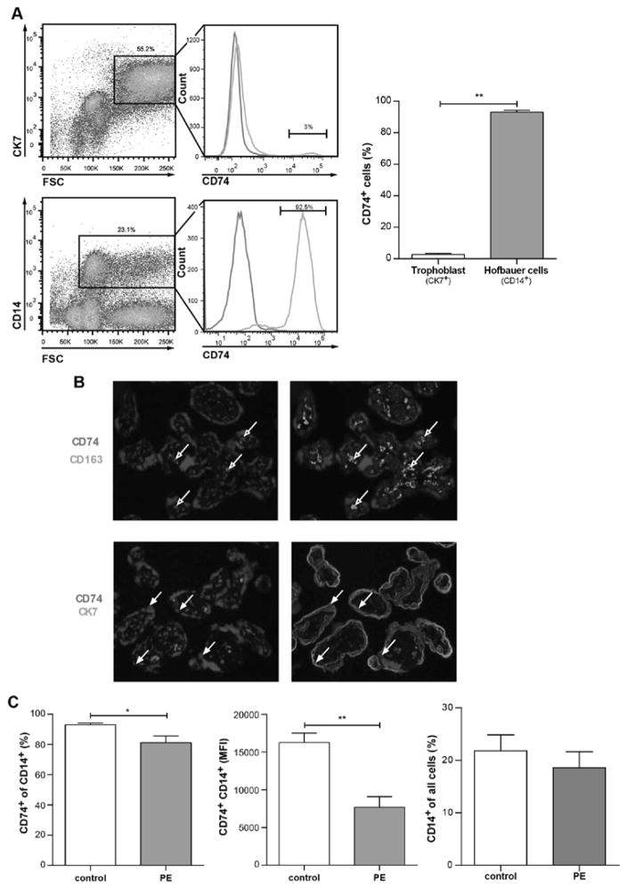
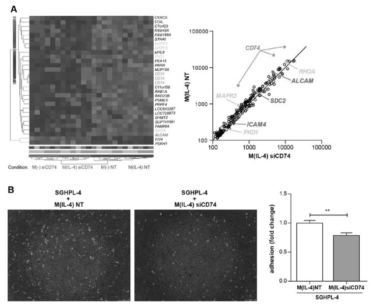
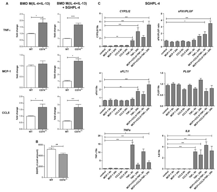
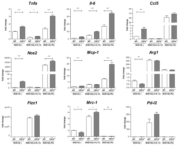
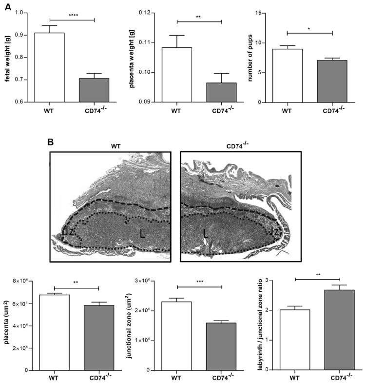
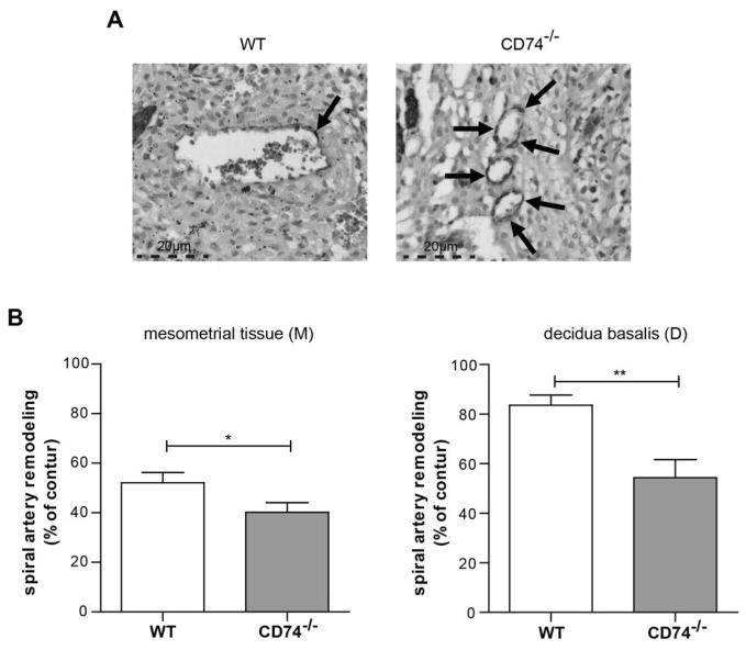
Methods and results: We performed whole-genome expression analysis of placental tissue. CD74 was one of the most downregulated genes in placentas from preeclamptic women. By reverse transcriptase-polymerase chain reaction, we confirmed this finding in early-onset (<34 gestational week, n=26) and late-onset (≥34 gestational week, n=24) samples from preeclamptic women, compared with healthy pregnant controls (n=28). CD74 protein levels were analyzed by Western blot and flow cytometry. We identified placental macrophages to express CD74 by immunofluorescence, flow cytometry, and RT-PCR. CD74-positive macrophages were significantly reduced in preeclamptic placentas compared with controls. CD74-silenced macrophages showed that the adhesion molecules ALCAM, ICAM4, and Syndecan-2, as well as macrophage adhesion to trophoblasts were diminished. Naive and activated macrophages lacking CD74 showed a shift toward a proinflammatory signature with an increased secretion of tumor necrosis factor-α, chemokine (C-C motif) ligand 5, and monocyte chemotactic protein-1, when cocultured with trophoblasts compared with control macrophages. Trophoblasts stimulated by these factors express more CYP2J2, sFlt1, TNFα, and IL-8. CD74-knockout mice showed disturbed placental morphology, reduced junctional zone, smaller placentas, and impaired spiral artery remodeling with fetal growth restriction.

Conclusions: CD74 downregulation in placental macrophages is present in preeclampsia. CD74 downregulation leads to altered macrophage activation toward a proinflammatory signature and a disturbed crosstalk with trophoblasts.

Keywords: immunology; macrophages; preeclampsia; pregnancy; trophoblasts.


Figures

Figure 1
Figure 1. CD74 is downregulated in preeclamptic placenta
A) CD74 expression is downregulated in placentas of preeclamptic women (PE) compared to healthy women (control; n=28). PE is subdivided in early onset PE (delivery <34 week of gestation; grey bar; n=26) and late onset PE (delivery ≥34 week of gestation; black bar; n=24) (**p<0.01; ***p<0.001; Bonferroni’s multiple comparisons test). B) CD74 protein (33 and 35 kDa) is lowered in placenta lysates of early and late onset PE (n=2 each). C) Confirmation by flow cytometry; representative CD74-positive gating is shown for whole placenta cell population (left panel). Mean fluorescent intensity (MFI) of CD74 is lowered in general placenta cell population of late PE vs. control (right panel) (n=10 (PE) and 6 (control); ***p=0.001; Mann Whitney test).
Figure 2
Figure 2. CD74 is highly expressed in placental macrophages (Hofbauer cells) and downregulated in preeclamptic Hofbauer cells
A) By flow cytometry; representative CK7-positive gating (trophoblast marker) and CD14-positve gating (macrophage marker) is shown for whole placenta cell population (left panel) of healthy controls. Solely 2.6±0.8 % of trophoblasts (CK7-positive) vs. 93.1±1.1 % of Hofbauer cells (CD14-positive) were positive for CD74 staining (right panel) (n=6; p<0.01; Mann Whitney test). B) Immunostaining showed that CD74 (red) is co-localized with CD163 (green) (Hofbauer cells; open arrows; upper panel) but not with CK7 (green). C) Flow cytometry on whole placenta cell population revealed that CD74 was less present in CD14-positive cells of placentas from late preeclamptic women (PE) compared to healthy women (control) (left panel). The mean fluorescent intensity (MFI) of CD74-CD14-positive cells was lower in PE vs. control (middle panel). Percentage of CD14-positive cells in all placental cells was not changed in PE (n=10) vs. control (n=6) (right panel) (*p<0.05, **p<0.01; Mann Whitney test).
Figure 3
Figure 3. Illustration of the maternal-fetal-interface
Tissue and cells from fetal origin are illustrated in blue. Maternal tissue and cells are illustrated in red. Fetal cytotrophoblasts invade into the maternal tissue and remodel the maternal spiral artery. Fetal macrophages (Hofbauer cells) interact with the fetal villous cytotrophoblast via adhesion molecules and cytokines.
Figure 4
Figure 4. Dysregulated expression and function of CD74 silenced M(−) and M(IL-4) macrophages
A) Heatmap represents microarray analysis for indicated conditions (left panel). Genes were pre-selected by 5% FDR (Subdued parametric three way ANOVA, Benjamini Hochberg FDR corrected) for CD74 silenced (siCD74) vs. non-targeting control (NT). Genes undergoing 65% FDR were plotted for siCD74 vs NT in M(IL-4) macrophages (right panel). Genes in orange correlate to “placenta” (by literature), genes in red encode adhesion molecules and CD74 is indicated in green (both panels). B) Adhesion of CD74 silenced macrophages (M(IL-4) siCD74) to first trimester trophoblast derived cell line (SGHPL-4) was lowered compared to control macrophages (M(IL-4) NT) as shown in representative pictures (left panel) and in summary of 3 independent experiments (right panel) (**p<0.01; unpaired t test).
Figure 5
Figure 5. Cytokine/chemokine-upregulation of macrophages derived from CD74 knockout mice (CD74−/−) and expression profile in a trophoblast cell line
A) In vitro activated bone marrow derived (BMD) macrophages (M(IL-4+IL-13)) from CD74−/− mice produced more tumor necrosis factor α (TNFα) and chemokine (C-C motif) ligand 5 (CCL5) compared to BMD M(IL-4+IL-13) from wild type (WT) mice (left panel) (n=6 each; *p<0.05, unpaired t test). Co-culture with human first trimester trophoblast derived cell line (SGHPL-4) enhanced the effect (right panel) (n=6 each, ***p<0.001, ****p<0.0001; unpaired t test). Secretion of monocyte chemoattractant protein-1 (MCP-1) was solely enhanced under co-culture conditions (n=4 each, *p<0.05, Mann Whitney test). All factors were secreted by macrophages, as Luminex assay was specific for mice. B) Co-culturing of BMD M(IL-4+IL-13) from CD74−/− with SGHPL-4 lowered cell amount of SGHPL-4 when compared to BMD M(IL-4+IL-13) from WT (n=30 each; **p<0.01, unpaired t test). C) SGHPL-4 stimulated by the combination of MCP-1, CCL5 and TNFα for 6h and 24h express more Cytochrome P450 Subfamily 2J Polypeptide 2 (CYP2J2), soluble Fms-like tyrosine kinase 1 (sFlt1), TNFα and Interleukin 8 (IL-8). The ratio of the expression of sFlt1 and the placental growth factor (PLGF), the sFlt1/PLGF ratio was also enhanced (n=9, *p<0.05, **p<0.01, ***p<0.001; ANOVA followed by Dunnett post hoc testing).
Figure 6
Figure 6. Pro-inflammatory signature of macrophages derived from CD74 knockout mice (CD74−/−)
In vitro differentiated bone marrow derived (BMD) macrophages (M(−)) from CD74−/− mice express more tumor necrosis factor α (Tnfα), interleukin 6 (Il-6), chemokine (C-C motif) ligand 5 (Ccl5), nitric oxide synthase 2 (Nos2) and monocyte chemoattractant protein-1 (Mcp-1) compared to BMD M(−) from wild type (WT) mice. Tnfα and Il-6 were also higher expressed in BMD M(IL-4+IL-13) from CD74−/− mice compared to WT. Marker for LPS signature (Nos2, Ccl5, IL-6, Tnfα, Mcp-1) and IL-4+IL-13 signature (arginase 1 (Arg1), Resistin-like molecule alpha1 (Fizz1), mannose receptor 1 (Mrc-1), programmed death ligand 2 (Pd-l2)) were fully activated in M(LPS) and M(IL-4+IL-13) respectively and pro-inflammatory activation by LPS was further increased by CD74−/− in regard of Tnfα, Il-6, Nos2 and Mcp-1 (n=6 each;*p<0.05, **p<0.01, ***p<0.001; unpaired t test).
Figure 7
Figure 7. Fetal growth restriction and disturbed placenta morphology in CD74 knockout mice (CD74−/−)
A) Pup and placental weights were lower in CD74−/− (n=32) vs. WT (n=33). Litter size was also decreased in CD74−/− (n=17) vs. WT (n=28) (*p<0.05, **p<0.01 and ****p<0.0001; Mann Whitney test). B) Representative pictures of Masson stained mid-sagittal placental tissue sections used to carry out the histomorphological analysis (upper panel). Zones are marked and indicated by L (labyrinth zone) and Jz (junctional zone). The overall size of the placenta was decreased in CD74−/− tissues (lower panel) whereas the ratio between the labyrinth/junctional zones was enhanced in CD74−/− (n=13) in comparison to WT (n=17). The area of the junctional zone of CD74−/− placentas was smaller compared to those of WT. (**p<0.01, ***p<0.001; unpaired t test).
Figure 8
Figure 8. Impaired spiral artery remodeling in CD74 knockout mice (CD74−/−)
A) α-actin staining is shown in representative pictures of arteries. Arrows are indicating α-actin stained VSMC’s. B) Spiral arteries of CD74−/− showed more α-actin in the vessel wall when compared to wild type (WT), indicating an impaired remodeling. Bar graphs show % of remodeling of artery (total loss of α-actin = 100%). (M): n=85 (WT), n=95 (CD74−/−); (D): n=36 (WT), n=24 (CD74−/−); *p<0,05, **p<0,01; unpaired t test.

References

    1. Hypertension in pregnancy. Report of the american college of obstetricians and gynecologists’ task force on hypertension in pregnancy. Obstetrics and gynecology. 2013;122:1122–1131. - PubMed
    1. Fayyad AM, Harrington KF. Prediction and prevention of preeclampsia and iugr. Early human development. 2005;81:865–876. - PubMed
    1. Sacks GP, Studena K, Sargent K, Redman CW. Normal pregnancy and preeclampsia both produce inflammatory changes in peripheral blood leukocytes akin to those of sepsis. Am J Obstet Gynecol. 1998;179:80–86. - PubMed
    1. Rusterholz C, Hahn S, Holzgreve W. Role of placentally produced inflammatory and regulatory cytokines in pregnancy and the etiology of preeclampsia. Semin Immunopathol. 2007;29:151–162. - PubMed
    1. Szarka A, Rigo J, Jr, Lazar L, Beko G, Molvarec A. Circulating cytokines, chemokines and adhesion molecules in normal pregnancy and preeclampsia determined by multiplex suspension array. BMC immunology. 2010;11:59. - PMC - PubMed

MeSH terms