. 2016 Jun 7;113(23):6526-31.
doi: 10.1073/pnas.1524292113. Epub 2016 May 19.

MerTK cleavage limits proresolving mediator biosynthesis and exacerbates tissue inflammation

Affiliations

MerTK cleavage limits proresolving mediator biosynthesis and exacerbates tissue inflammation

Bishuang Cai et al. Proc Natl Acad Sci U S A. .

Abstract

The acute inflammatory response requires a coordinated resolution program to prevent excessive inflammation, repair collateral damage, and restore tissue homeostasis, and failure of this response contributes to the pathology of numerous chronic inflammatory diseases. Resolution is mediated in part by long-chain fatty acid-derived lipid mediators called specialized proresolving mediators (SPMs). However, how SPMs are regulated during the inflammatory response, and how this process goes awry in inflammatory diseases, are poorly understood. We now show that signaling through the Mer proto-oncogene tyrosine kinase (MerTK) receptor in cultured macrophages and in sterile inflammation in vivo promotes SPM biosynthesis by a mechanism involving an increase in the cytoplasmic:nuclear ratio of a key SPM biosynthetic enzyme, 5-lipoxygenase. This action of MerTK is linked to the resolution of sterile peritonitis and, after ischemia-reperfusion (I/R) injury, to increased circulating SPMs and decreased remote organ inflammation. MerTK is susceptible to ADAM metallopeptidase domain 17 (ADAM17)-mediated cell-surface cleavage under inflammatory conditions, but the functional significance is not known. We show here that SPM biosynthesis is increased and inflammation resolution is improved in a new mouse model in which endogenous MerTK was replaced with a genetically engineered variant that is cleavage-resistant (Mertk(CR)). Mertk(CR) mice also have increased circulating levels of SPMs and less lung injury after I/R. Thus, MerTK cleavage during inflammation limits SPM biosynthesis and the resolution response. These findings contribute to our understanding of how SPM synthesis is regulated during the inflammatory response and suggest new therapeutic avenues to boost resolution in settings where defective resolution promotes disease progression.

Keywords: 5-lipoxygenase; MerTK; efferocytosis; inflammation resolution; macrophages.


Conflict of interest statement

The authors declare no conflict of interest.

Figures

Fig. 1.
Fig. 1.
MerTK deficiency delays resolution, decreases PMN efferocytosis, and lowers SPM levels in sterile peritonitis. (A) WT or Mertk−/− mice were injected intraperitoneally with 1 mg of zymosan per mouse, and PMNs in the exudate were counted at the indicated times after zymosan injection. (B) Exudate leukocytes at the 30-h time point were costained with annexin V (apoptosis) and anti-Ly6G antibody (PMNs), and the percent of annexin V+ PMNs was detected by flow cytometry. (C) To assess efferocytosis, 30-h exudate leukocytes were first stained with anti-F4/80 antibody, permeabilized and stained for Ly6G, and then analyzed by flow cytometry for F4/80+Ly6G+ cells among total F4/80+ cells. Mϕs, macrophages. (D) Exudates were assayed for relative levels of immunoreactive (i) SPMs by LXA4 and RvD1 ELISAs, noted as LX-i and RvD-i, respectively. (E) BMDMs were treated for 1 h with MerTK-activating antibody (activ-αMer) or control IgG, and the media were assayed for relative LX-i and RvD-i, as described in D. (F) Human monocyte-derived macrophages were treated with scrambled (Scr) RNA or siRNA against Mertk and then treated for 1 h with vehicle control or human Gas6. The media were assayed for relative LX-i and RvD-i as in D. *P < 0.05 [vs. WT (mean ± SEM, n = 4 mice per group; AD); and vs. IgG or vehicle control (mean ± SEM, n = 3 experiments; E and F)]; n.s., not significant.
Fig. S1.
Fig. S1.
FACS gating strategy for assessing apoptotic PMNs and efferocytosis in the exudates of zymosan-treated mice. (A and C) Apoptotic PMNs were assessed by costaining with Annexin V and Ly6G. (B and D) Apoptotic PMNs engulfed by macrophages were assessed by costaining with F4/80 and Ly6G, as described in Fig. 1 B and C.
Fig. S2.
Fig. S2.
LC-MS/MS analysis of lipid mediators in WT, Mertk−/−, and MertkCR mice. (A) PLS-DA 2D score plot of lipid mediators measured in peritoneal exudates of WT, Mertk−/−, and MertkCR mice 20 h after zymosan administration. (BD) Levels of SPMs (RvD1-6, RvE1, RvE3, LXA4, 15-epi-LXA4, LXB4, 15-epi-LXB4, LXB4 isomer, Mar2, 17R-PD1 and 10S,17S-diHDHA), prostaglandins (PGD2, PGE2, and PGF), and LTB4 in peritoneal exudates 20 h after zymosan administration. Data are mean ± SEM; n = 5 mice per genotype. *P < 0.05 vs. WT by one-way ANOVA, followed by Dunnett’s posttests; ns, not significant.
Fig. S3.
Fig. S3.
Exudate TGF-β is decreased and TNF-α is increased in Mertk−/− mice, with an opposite effect in MertkCR mice. (A and C) WT, Mertk−/−, or MertkCR mice were injected intraperitoneally with 1 mg of zymosan per mouse. After 30 h, exudate fluid was harvested and subjected to TGF-β and TNF-α measurement by ELISA. (B) Expression of cell-surface CD206 and cellular iNOS expression in 30-h exudate F4/80+ macrophages from WT, Mertk−/−, or MertkCR mice was quantified by flow-cytometric analysis.
Fig. S4.
Fig. S4.
Validation of activating anti-MerTK antibody and Mertk siRNA, and LC-MS/MS analysis of lipid mediators in WT and Mertk−/− macrophages. (A) BMDMs were incubated for 20 min with 5 nM MerTK-activating antibody (activ-αMer) or control IgG and then assayed for phospho- and total MerTK by immunoblot. (B) Human monocyte-derived macrophages were treated with scrambled (Scr) RNA control or siRNA against Mertk and then assayed for MerTK by immunoblot. (C) Bone marrow-derived WT or Mertk−/− murine macrophages treated for 1 h with vehicle control (Veh) or human Gas6 and then assayed by LC-MS/MS for DHA-derived SPMs (RvD1-6, 10S,17S-diHDHDA, 17R-PD1, and their biosynthetic pathway markers, 17-, 7-, and 4-HDHA) and AA-derived SPMs (LXA4, 15-epi-LXA4, LXB4, 15-epi-LXB4, LXB4 isomer, and their biosynthetic pathway marker, 15-HETE). *P < 0.05 vs. vehicle control; #P < 0.05 between Gas6-treated WT and Mertk−/− macrophages (mean ± SEM, n = 3/group). (D) BMDMs were incubated with Gas6 for 1 h and then assayed by quantitative RT-PCR for Hpgd (15-PGDH) mRNA relative to 36B4 mRNA.
Fig. S5.
Fig. S5.
Design of MertkCR mouse. Diagram shows design of the targeting alleles used to make the MertkCR mouse (see SI Materials and Methods for details). The targeting vector contained a mutated version of exon 10 of the Mertk gene in which the codons encoding residues 483–488 of MerTK were deleted and a downstream Neo cassette (NeoR) flanked by LoxP sites. ES clones containing this vector via homologous recombination were used to generate germ-line chimeras, which were then bred to EIIa-Cre mice to remove the NEO cassette. (Upper gel): Representative panel from PCR screening of 96 ES clones, using primer set 1, to identify the targeted Neo-containing Mertk mutant allele. L = 1 kb plus DNA ladder; ES clone b shows an ES cell clone with the predicted 2,719-bp amplicon for the targeting vector. (Lower gel): Representative panel of PCR screen, using primer set 2, of DNA from 18 pups resulting from the cross of male chimeras crossed to EIIa (cre/cre) females. The WT amplicon is 348 bp (lower band), and the targeted Neo-minus Mertk mutant allele (L83) is 431 bp (upper band). A total of three pups were found to carry the L83 locus, including pup 6 as shown. L = Invitrogen 1 kb plus ladder.
Fig. S6.
Fig. S6.
Macrophage cell-surface MerTK and peripheral immune cells are similar in WT and MertkCR mice. (A) Resident peritoneal cells from WT and MertkCR mice were immunostained for F4/80 and Ly6G and then analyzed by flow cytometry. (B) The mean fluorescence intensity (MFI) of cell-surface MerTK on F4/80+ cells was quantified by flow cytometry (mean ± SEM, n = 4 mice per group; n.s., nonsignificant). (C and D) WT and MertkCR blood and splenic cells were immunostained for Ly6G, CD115, CD3, and B220 antibodies and then analyzed by flow cytometry to detect PMNs, monocytes, T cells, and B cells, respectively.
Fig. 2.
Fig. 2.
MertkCR macrophages are resistant to cleavage and maintain efferocytosis under inflammatory conditions in vitro. (A and B) BMDMs from a WT mouse or two different MertkCR mice were incubated for 2 h with 50 ng/mL LPS or vehicle control (Veh). The media were assayed for sol-Mer by immunoblot (A), and the cells were assayed for cell-surface MerTK by flow cytometry (B); flow data and quantified mean fluorescence intensity (MFI) data are shown. (C) BMDMs were treated as in A and then incubated for 1 h with Calcein Green-labeled apoptotic Jurkat cells. The percent of total BMDMs with internalized labeled ACs was quantified by analysis of fluorescence microscopy images. *P < 0.001 (vs. Veh; mean ± SEM, n = 3 experiments); n.s., not significant.
Fig. S7.
Fig. S7.
MertkCR macrophages respond normally to LPS and are resistant to PMA-induced MerTK cleavage. (A) BMDMs from WT or MertkCR mice were treated with 50 ng/mL LPS for the indicated times and then assayed by immunoblot for phospho- and total NF-κB and P38. (B) BMDMs from WT or MertkCR mice were treated with 50 nM PMA or vehicle control for 2 h, and the media were then assayed for sol-Mer by immunoblot. (C) As in B, but the cells were analyzed for cell-surface MerTK by flow cytometry. *P < 0.001 vs. all other groups (mean ± SEM, n = 3 experiments); n.s., nonsignificant.
Fig. 3.
Fig. 3.
Resolution of sterile peritonitis is improved in MertkCR mice. (A) WT or MertkCR mice were injected intraperitoneally with zymosan as in Fig 1, and exudate PMN were quantified at the indicated times after zymosan injection. (B) A portion of the peritoneal exudate harvested at 30 h was assayed for sol-Mer by immunoblot, and another portion was costained with anti-F4/80 and -MerTK, followed by flow-cytometric quantification of cell-surface MerTK on F4/80+ cells. (C and D) Apoptotic PMNs and efferocytosis of PMNs by macrophages in the 30-h exudate were determined as in Fig. 1 B and C. (E) Relative levels of exudate LX-i and RvD-i were measured at 30 h by ELISA. (G) BMDMs were treated with 50 nM PMA or vehicle control for 2 h. The cells were then incubated for 1 h with MerTK-activating antibody (activ-αMer) or IgG, and the media were assayed for relative LX-i by ELISA. (G) As in A, but at 20 h after zymosan, the mice were injected intraperitoneally with blocking antibody against ALX or IgG control, and then exudate PMNs were quantified at 30 h after zymosan. *P < 0.05 [vs. WT (mean ± SEM, n = 4 mice per group; AE)]. In F, bars with asterisks are statistically the same among each other and different from all other bars at P < 0.05 (mean ± SEM, n = 3 experiments); for G, different symbols indicate values that are statistically different from each other and from IgG control at P < 0.05 (mean ± SEM, n = 4 mice per group).
Fig. S8.
Fig. S8.
PMNs do not express MerTK. WT mice were challenged with 1 mg of zymosan intraperitoneally After 4 h, PMNs from either peritoneal exudates or blood were isolated and purified with a PMN purification kit (Miltenyl Biotec). MerTK from purified PMNs was detected by immunoblotting (BMDMs serve as a positive control).
Fig. 4.
Fig. 4.
I/R-induced lung damage is increased in Mertk−/− mice and decreased in MertkCR mice, with reciprocal changes in plasma LX-i. Mertk−/−, MertkCR, or littermate WT mice were subjected to 60-min bilateral hindlimb ischemia followed by 90-min reperfusion. (A) Lung extracts were assayed for MPO content by ELISA (sham values for WT, Mertk−/−, and MertkCR were 6.2, 7.7, and 5.5 pg/mg protein, respectively). (B) Perfused and fixed lungs were scored for lung injury as described in Materials and Methods. Representative images are shown in Fig. S9. Sham values for WT, Mertk−/−, and MertkCR were 0.17, 0.176, and 0.148, respectively. (C) Plasma was assayed for relative LX-i by ELISA (sham values for WT, Mertk−/−, and MertkCR were 137.5, 108.8, and 106.2 pg/mL, respectively). (D) Whole blood was immunostained for CD45, Gr1, and CD41, and the percent of Gr1+CD41+ cells (PMN-platelet aggregates) among total CD45+ cells was quantified by flow cytometry (sham values for WT, Mertk−/−, and MertkCR were 1.5%, 1.3%, and 1.6%, respectively). *P < 0.002 (mean ± SEM, n = 6–10 mice per group).
Fig. S9.
Fig. S9.
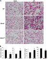
Hindlimb I/R-induced lung injury is greater in Mertk−/− mice and less in MertkCR mice compared with WT mice. (A) WT, Mertk−/−, and MertkCR mice were subjected to 60-min bilateral hindlimb ischemia followed by 120-min reperfusion. Perfused and fixed lungs were then sectioned and stained with hematoxylin and eosin. The quantified lung injury score data appear in Fig. 4B. Arrows, neutrophils. (Magnification, 20×.) (B) After I/R, plasma TGF-β and TNF-α from WT, Mertk−/−, and MertkCR mice were measured by ELISA. *P < 0.05 vs. WT control mice.
Fig. 5.
Fig. 5.
MerTK promotes SPM production via an increase in the cytoplasmic:nuclear ratio of 5-LOX. (A) BMDMs were treated for 1 h with activ-αMer or control IgG. The cells were then fixed, permeabilized, and immunostained for 5-LOX, with nuclear DAPI costain. (Scale bar, 10 μm.) The cytoplasmic:nuclear ratio of 5-LOX was quantified from confocal immunofluorescence images. (B) Similar to A, except apoptotic Jurkat cells were used instead of activating antibody. (C) Human monocyte-derived macrophages were treated for 1 h with vehicle control (Veh) or human Gas6 and then immunoblotted for p-Ser271–5-LOX and total 5-LOX. (D) WT, Mertk−/−, or MertkCR mice were injected intraperitoneally with 1 mg of zymosan per mouse, and 30 h later, exudate macrophages were assayed for cytoplasmic:nuclear ratio of 5-LOX. (E) BMDMs from Alox5−/− mice were transfected with plasmids encoding WT 5-LOX or S271D–5-LOX and then incubated with MerTK-activating antibody for 1 h. The cytoplasmic:nuclear ratio of 5-LOX was quantified from confocal immunofluorescence images. (F) BMDMs from Alox5−/− mice were transfected with plasmids encoding WT 5-LOX or S271D–5-LOX and then incubated with activ-αMer for 1 h. The media were assayed for relative LX-i by ELISA. (A and B) *P < 0.05 vs. IgG (mean ± SEM, n = 3 experiments); (D) #P < 0.05 vs. WT (mean ± SEM, n = 4 mice/group); and (E) *P < 0.05 vs. all other groups (mean ± SEM, n = 3 experiments).
Fig. S10.
Fig. S10.
TGFβ signaling is not involved in MerTK-mediated LX-i synthesis, and MerTK activation reduces phospho-MK2 and enhances cytosol 5-LOX in human monocyte-derived macrophages. (A) BMDMs were pretreated with either vehicle or 10 µM LY2109761 (TGF-β receptor inhibitor) for 30 min. Cells were then incubated with either IgG or activ-αMer for 1 h, and the media were collected and assayed for LX-i by ELISA. n.s., nonsignificant. (A, Inset) p-SMAD2 immunoblot as a positive control for the inhibitory activity of LY2109761. (B and C) Human monocyte-derived macrophages were treated for 1 h with vehicle control or human Gas6 and then either immunoblotted for p-MK2 and total MK2 or fractionated into cytosolic and nuclear fractions and immunoblotted for 5-LOX, the nuclear protein lamin A/C, and the cytosolic protein GAPDH.

References

    1. Serhan CN. Pro-resolving lipid mediators are leads for resolution physiology. Nature. 2014;510(7503):92–101. - PMC - PubMed
    1. Medzhitov R. Inflammation 2010: New adventures of an old flame. Cell. 2010;140(6):771–776. - PubMed
    1. Nathan C, Ding A. Nonresolving inflammation. Cell. 2010;140(6):871–882. - PubMed
    1. Tabas I, Glass CK. Anti-inflammatory therapy in chronic disease: Challenges and opportunities. Science. 2013;339(6116):166–172. - PMC - PubMed
    1. Viola J, Soehnlein O. Atherosclerosis—A matter of unresolved inflammation. Semin Immunol. 2015;27(3):184–193. - PubMed

Publication types

MeSH terms