

Simultaneous Segmentation of Retinal Surfaces and Microcystic Macular Edema in SDOCT Volumes
- PMID: 27199502
- PMCID: PMC4869874
- DOI: 10.1117/12.2214676
Simultaneous Segmentation of Retinal Surfaces and Microcystic Macular Edema in SDOCT Volumes
Abstract
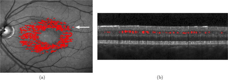
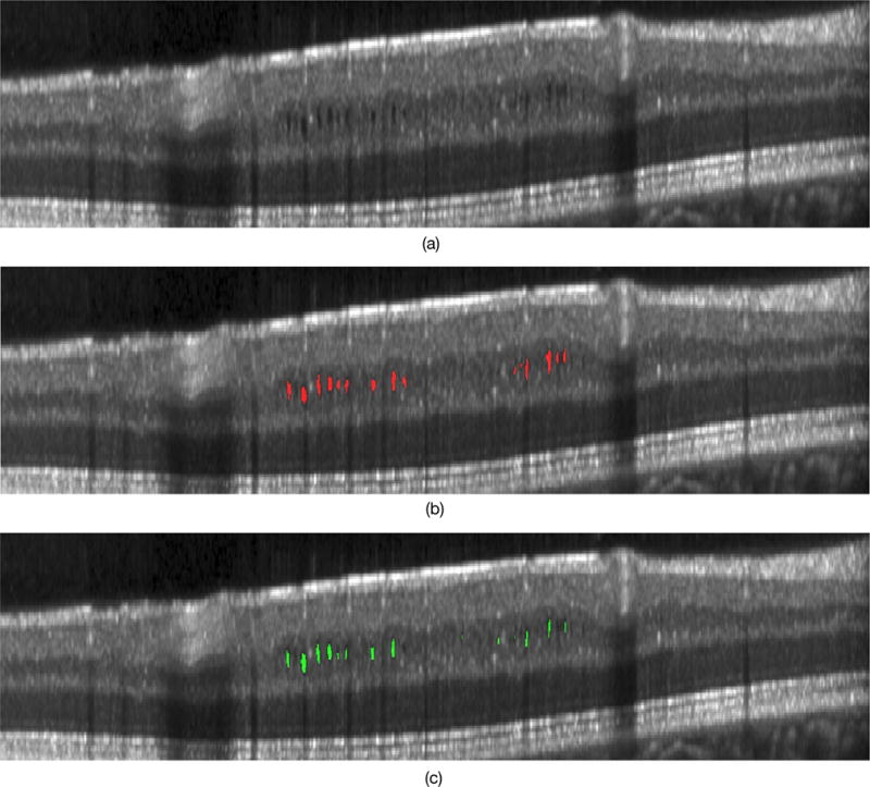
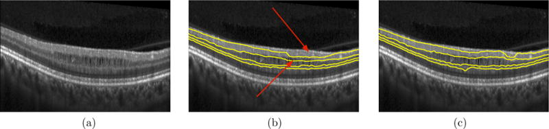
Optical coherence tomography (OCT) is a noninvasive imaging modality that has begun to find widespread use in retinal imaging for the detection of a variety of ocular diseases. In addition to structural changes in the form of altered retinal layer thicknesses, pathological conditions may also cause the formation of edema within the retina. In multiple sclerosis, for instance, the nerve fiber and ganglion cell layers are known to thin. Additionally, the formation of pseudocysts called microcystic macular edema (MME) have also been observed in the eyes of about 5% of MS patients, and its presence has been shown to be correlated with disease severity. Previously, we proposed separate algorithms for the segmentation of retinal layers and MME, but since MME mainly occurs within specific regions of the retina, a simultaneous approach is advantageous. In this work, we propose an automated globally optimal graph-theoretic approach that simultaneously segments the retinal layers and the MME in volumetric OCT scans. SD-OCT scans from one eye of 12 MS patients with known MME and 8 healthy controls were acquired and the pseudocysts manually traced. The overall precision and recall of the pseudocyst detection was found to be 86.0% and 79.5%, respectively.
Keywords: graph-cuts; graph-theoretic approach; microcysts; multiple surface segmentation; optical coherence tomography; retina.
Figures
Similar articles
-
Segmentation of microcystic macular edema in Cirrus OCT scans with an exploratory longitudinal study.Proc SPIE Int Soc Opt Eng. 2015;9417:94170P. doi: 10.1117/12.2082164. Proc SPIE Int Soc Opt Eng. 2015. PMID: 26023249 Free PMC article.
-
Deep learning based topology guaranteed surface and MME segmentation of multiple sclerosis subjects from retinal OCT.Biomed Opt Express. 2019 Sep 12;10(10):5042-5058. doi: 10.1364/BOE.10.005042. eCollection 2019 Oct 1. Biomed Opt Express. 2019. PMID: 31646029 Free PMC article.
-
The clinical spectrum of microcystic macular edema.Invest Ophthalmol Vis Sci. 2014 Feb 18;55(2):952-61. doi: 10.1167/iovs.13-12912. Invest Ophthalmol Vis Sci. 2014. PMID: 24398089
-
Automatic Anisotropic Diffusion Filtering and Graph-search Segmentation of Macular Spectral-domain Optical Coherence Tomographic (SD-OCT) Images.Curr Med Imaging Rev. 2019;15(3):308-318. doi: 10.2174/1573405613666171201155119. Curr Med Imaging Rev. 2019. PMID: 31989882 Review.
-
Microcystic macular oedema in optic neuropathy: case series and literature review.Clin Exp Ophthalmol. 2018 Dec;46(9):1075-1086. doi: 10.1111/ceo.13327. Epub 2018 Jun 20. Clin Exp Ophthalmol. 2018. PMID: 29799159 Review.
Cited by
-
Deep-learning approach for automated thickness measurement of epithelial tissue and scab using optical coherence tomography.J Biomed Opt. 2022 Jan;27(1):015002. doi: 10.1117/1.JBO.27.1.015002. J Biomed Opt. 2022. PMID: 35043611 Free PMC article.
-
Structured layer surface segmentation for retina OCT using fully convolutional regression networks.Med Image Anal. 2021 Feb;68:101856. doi: 10.1016/j.media.2020.101856. Epub 2020 Oct 14. Med Image Anal. 2021. PMID: 33260113 Free PMC article.
-
Longitudinal deep network for consistent OCT layer segmentation.Biomed Opt Express. 2023 Apr 3;14(5):1874-1893. doi: 10.1364/BOE.487518. eCollection 2023 May 1. Biomed Opt Express. 2023. PMID: 37206119 Free PMC article.
-
Layer boundary evolution method for macular OCT layer segmentation.Biomed Opt Express. 2019 Feb 4;10(3):1064-1080. doi: 10.1364/BOE.10.001064. eCollection 2019 Mar 1. Biomed Opt Express. 2019. PMID: 30891330 Free PMC article.
-
Segmentation of mouse skin layers in optical coherence tomography image data using deep convolutional neural networks.Biomed Opt Express. 2019 Jun 21;10(7):3484-3496. doi: 10.1364/BOE.10.003484. eCollection 2019 Jul 1. Biomed Opt Express. 2019. PMID: 31467791 Free PMC article.
References
-
- de Boer JF, Cense B, Park BH, Pierce MC, Tearney GJ, Bouma BE. Improved signal-to-noise ratio in spectral-domain compared with time-domain optical coherence tomography. Opt Lett. 2003;28(21):2067–2069. - PubMed
-
- Ratchford JN, Saidha S, Sotirchos ES, Oh JA, Seigo MA, Eckstein C, Durbin MK, Oakley JD, Meyer SA, Conger A, Frohman TC, Newsome SD, Balcer LJ, Frohman EM, Calabresi PA. Active MS is associated with accelerated retinal ganglion cell/inner plexiform layer thinning. Neurology. 2013;80(1):47–54. - PMC - PubMed
-
- Saidha S, Sotirchos ES, Ibrahim MA, Crainiceanu CM, Gelfand JM, Sepah YJ, Ratchford JN, Oh J, Seigo MA, Newsome SD, Balcer LJ, Frohman EM, Green AJ, Nguyen QD, Calabresi PA. Microcystic macular oedema, thickness of the inner nuclear layer of the retina, and disease characteristics in multiple sclerosis: a retrospective study. Lancet Neurol. 2012;11(11):963–972. - PMC - PubMed
-
- Burggraaff MC, Trieu J, de Vries-Knoppert WAEJ, Balk L, Petzold A. The clinical spectrum of microcystic macular edema. Investig Ophthalmol Vis Sci. 2014;55(2):952–961. - PubMed
Grants and funding
LinkOut - more resources
Full Text Sources
Other Literature Sources
Miscellaneous