

Muscle MRI in muscular dystrophies
Acta Myol.
2015 Dec.
Abstract
Muscle MRI has become a very useful tool in the diagnosis and follow-up of patients with muscle dystrophies. Muscle MRI provides us about many aspects of the structure and function of skeletal muscles, such as the presence of oedema or fatty infiltration. In the last years many reports have described the particular muscles that are involved in these muscle disease. This knowledge can facilitate the diagnosis in many cases. In the present paper we review the main changes observed in muscle MRI of patients with muscle dystrophies.
Keywords: Limb-girdle muscular dystrophies; muscle MRI.
Figures
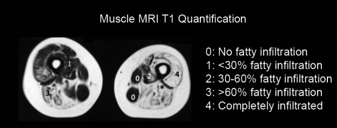
Diagram showing our quantification method of the fatty infiltration present in T1 sequences. In the image, a thigh slice is quantified using the Mercuri scale modified by Fischer.
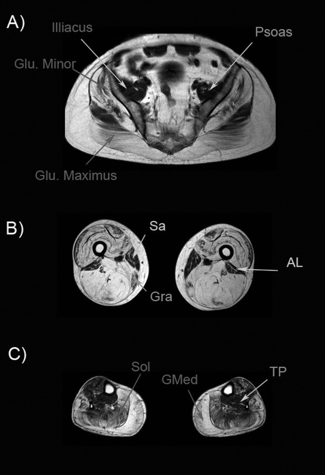
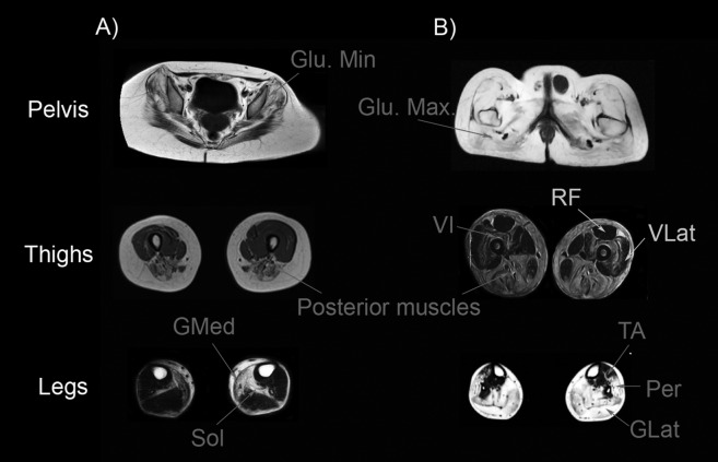
Muscle MRI of a Becker muscle dystrophy patient. A) Pelvic image shows atrophy of glutei muscles (Gluteus minimus and maximus are shown in the image). Psoas and illiacus muscle are usually spared until late stages of the disease. B) Image of the thigh shows a complete atrophy of all muscles, except for sartorius (Sar), gracillis (Gra) and adductor longus (AL). C) Image of the legs showing a severe atrophy of gastrocnemius medialis (GMed) and soleus (Sol). In this patient, tibialis posterior (TP) was not involved.
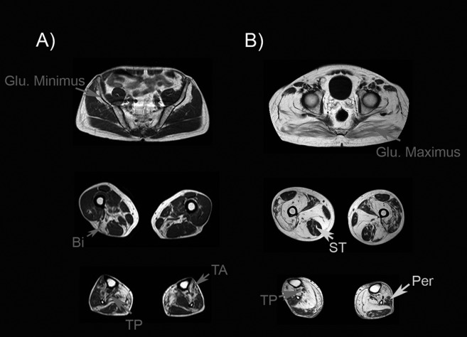
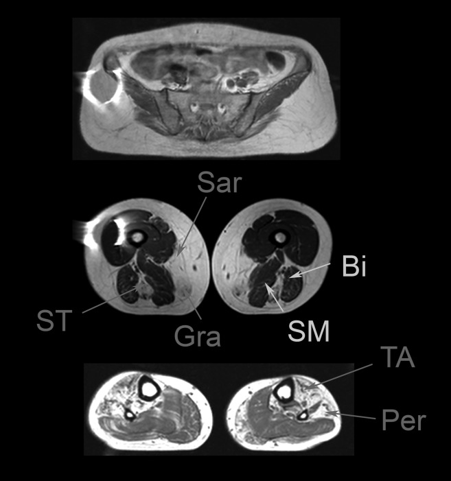
Muscle MRI of two patients with mutations in the MYOT gene. A). Patient with mild weakness: muscle MRI shows mild involvement of gluteus minimus, biceps (Bi), tibialis anterior (TA) and tibialis posterior (TP). B) Patient with severe weakness: muscle MRI shows involvement of all glutei muscles, although gluteus maximus is less involved than gluteus minimus or medius. In the thighs, semitendinosus (ST) is not involved. In contrast, there is a clear involvement of the posterior muscles of the thighs. Tibialis posterior (TP) is more atrophic than peroneus muscle (Per).
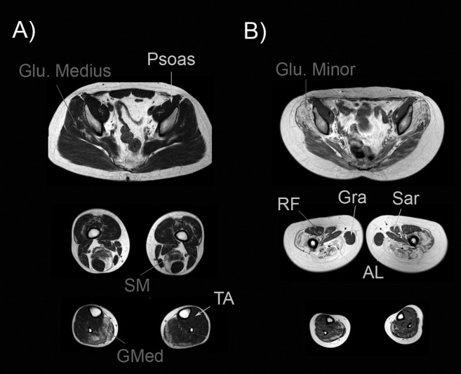
Muscle MRI of two patients with mutations in the LMNA gene. A) Patient with mild weakness. In this patient we observed fatty infiltration in the gluteus medius, semimembranosus (SM) and gastrocnemius medialis (GMed) muscles. B) Patient with severe weakness: fatty infiltration was observed in gluteus minimus and medius. All the muscles of the thighs were involved, except rectus femoris (RF), adductor longus (AL), sartorius (Sar) and gracillis (Gra) that were not atrophic.
Muscle MRI of a patient with mutation is the DES gene. Muscle MRI shows a preferential involvement of semitendinosus (ST), sartorius (Sar) and gracillis (Gra) muscles, while in contrast semimembranosus (SM) and biceps (Bi) are not involved. In the legs, there is an involvement of tibialis anterior (TA) and peroneus muscles (Per). Courtesy of Dr. Giorgio Tasca.
Muscle MRI in patients with mutations in the CAPN3 gene. Gluteus minimus and medius tend to be more involved than gluteus maximus (A) in most of the patients, although in advanced stages, all glutei muscles can be completely atrophic. In general, posterior muscles of the thigh are more severely involved than anterior muscles (A), but is not uncommon to find involvement of vasti muscles, especially vastus intermedius (VI). Atrophy of gastrocnemius medialis (GMed) and soleus (Sol) is frequently found (A), but in advanced cases (B) is not uncommon to find atrophy of gastrocnemius lateralis (GLat), peroneus (Per) and tibialis anterior (TA) muscles.
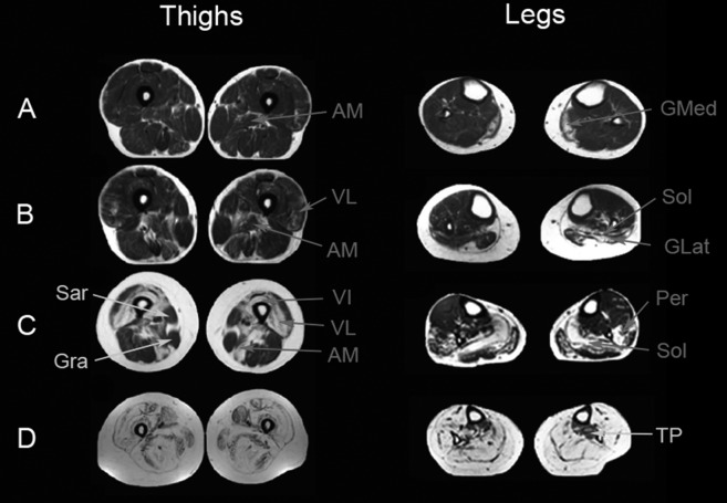
Progression of muscle atrophy in patients with mutations in the DYSF gene. There is a mild involvement of the adductor major (AM) and gastrocnemius medialis (GMed) in the first stages of the disease (A). In a second stage (B), atrophy involves also the vastus lateralis (VL) and the soleus (Sol). Then (C), fatty infiltration progresses and involves the rest of the vasti muscles and the posterior muscles of the thighs, but sartorius (sar) and gracillis (gra) are commonly not involved. In the legs, the vastus lateralis and peroneus become atrophic. In the most advanced stages (D), all the muscles are infiltrated by fat, but in some patients tibialis posterior (TP) may be not involved.
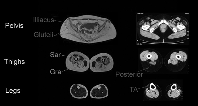
Muscle MRI of patients with sarcoglycanopathy and titinopathy. A: 35 years old patient with LGMD-2D: there is a severe involvement of pelvic muscles (including glutei and psoas muscles) and thigh muscles. Sartorius (sar) and gracillis (gra) are not involved until advanced stages of the diseases. In contrast leg muscles are not involved. B: Muscle CT of a 62 years old patient with distal titinopathy: preferential involvement of the tibialis anterior (TA) and the posterior muscles of the thighs is observed. In contrast, the pelvis muscles are not involved.
Muscle MRI of a patient with mutations in the ANO5 gene. A: Muscle MRI of a 53 years old patient with mild involvement of the lower limbs. Axial images showed no involvement of pelvic muscles, but in contrast there was a clear asymmetric involvement of vastus lateralis (VL) and posterior muscles of the thighs. In the legs, muscles of the posterior compartment were also involved, including soleus (Sol) and gastrocnemius medialis. B and C: hypenintensities in STIR sequences were observed in the posterior muscles of the thighs and in the vastus medialis muscle (arrows).
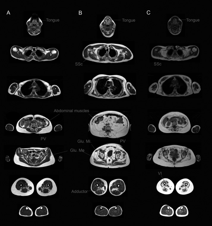
Muscle MRI of patients with Pompe disease. A: Whole body muscle MRI of a 35 years olf patient with mild muscle involvement showing atrophy of tongue, paravertebral and abdominal muscles, gluteus medius (Glu.Me) and posterior muscles of the thighs. B: Muscle MRI of a 43 years old patients with moderate weakness showing involvement of tongue, subscapularis (SSc), paravertebral and abdominal muscles, gluteus medius (Glu.Me) and gluteus minimus and adductor major (AM). C: Muscle MRI of a 44 years old patients with severe weakness showing involvement of tongue, subscapularis (SSc), paravertebral, abdominal, all glutei muscles and anterior and posterior muscles of the thighs.
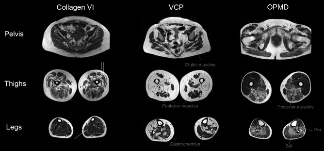
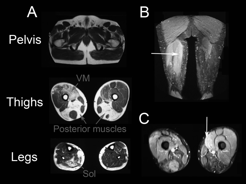
Muscle MRI of patients with collagen VI related myopathy, HIBMPD and OPMD muscle dystrophies. A: Muscle MRI of a 20 years old patient with mutations in the COL6A3 gene showing concentric atrophy of the vastus lateralis (double arrow) and a band of atrophy between gastrocnemius medialis and soleus (single arrow). B: Muscle MRI of a 56 years old woman with a mutation in the VCP gene producing patchy atrophy of the glutei, posterior muscles of the thighs and both gastrocnemius. C: Muscle MRI of a 72 years old patient with OPMD showing preferential involvement of posterior muscles of the thighs and the soleus muscle.
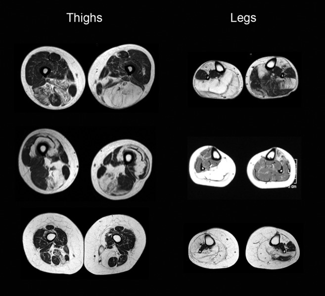
Muscle MRI of patients with Facio-Scapulo-Humeral muscle dystrophy. Muscle MRI of different patients with FSH muscle dystrophy showing asymmetries in the thighs and legs studies.
References
-
- Carlier PG, Mercuri E, Straub V. Applications of MRI in muscle diseases. Neuromuscul Disord. 2012;22(Suppl 2):S41–S41. - PubMed
-
- Paradas C, Llauger J, Diaz-Manera J, et al. Redefining dysferlinopathy phenotypes based on clinical findings and muscle imaging studies. Neurology. 2010;75:316–323. - PubMed
-
- Dahlqvist JR, Vissing CR, Thomsen C, Vissing J. Severe paraspinal muscle involvement in facioscapulohumeral muscular dystrophy. Neurology. 2014;83:1178–1183. - PubMed
-
- Tasca G, Iannaccone E, Monforte M, et al. Muscle MRI in Becker muscular dystrophy. Neuromuscul Disord. 2012;22(Suppl 2):S100–S106. - PubMed
MeSH terms
LinkOut - more resources
Full Text Sources
Other Literature Sources
Medical