. 2016 Apr 25:10:31.
doi: 10.3389/fncir.2016.00031. eCollection 2016.

Phasic and Tonic mGlu7 Receptor Activity Modulates the Thalamocortical Network

Affiliations

Phasic and Tonic mGlu7 Receptor Activity Modulates the Thalamocortical Network

Valériane Tassin et al. Front Neural Circuits. .

Abstract

Mutation of the metabotropic glutamate receptor type 7 (mGlu7) induces absence-like epileptic seizures, but its precise role in the somatosensory thalamocortical network remains unknown. By combining electrophysiological recordings, optogenetics, and pharmacology, we dissected the contribution of the mGlu7 receptor at mouse thalamic synapses. We found that mGlu7 is functionally expressed at both glutamatergic and GABAergic synapses, where it can inhibit neurotransmission and regulate short-term plasticity. These effects depend on the PDZ-ligand of the receptor, as they are lost in mutant mice. Interestingly, the very low affinity of mGlu7 receptors for glutamate raises the question of how it can be activated, namely at GABAergic synapses and in basal conditions. Inactivation of the receptor activity with the mGlu7 negative allosteric modulator (NAM), ADX71743, enhances thalamic synaptic transmission. In vivo administration of the NAM induces a lethargic state with spindle and/or spike-and-wave discharges accompanied by a behavioral arrest typical of absence epileptic seizures. This provides evidence for mGlu7 receptor-mediated tonic modulation of a physiological function in vivo preventing synchronous and potentially pathological oscillations.

Keywords: EEG; epilepsy; glutamate; short-term plasticity; thalamic network.


Figures

FIGURE 1
FIGURE 1
The mGlu7 receptor modulates glutamatergic transmission at VPM synapses onto TRN neurons. (A) Schematic representation of the thalamocortical and intra-thalamic connections between glutamatergic and GABAergic neurons in the somatosensory cortex (S1), the thalamic reticular nucleus (TRN) composed exclusively of GABAergic neurons, and the ventro-postero-medial nucleus (VPM), devoid of interneurons. FS, Fast-Spiking interneurons; RS, Regular-Spiking interneurons; Exc, excitatory glutamatergic neurons. (B) Experimental configuration, including the positions of the electrical stimulation into the VPM and the recording electrode into the TRN. (C) Single and average amplitudes of evoked EPSCs in WT and mGlu7aAAA mice. (D,F) Representative evoked EPSCs recorded from TRN neurons in response to VPM stimulation of WT (left) and mGlu7aAAA (right) mice and effect of D,L-AP4 (1.2 mM, D) and ADX71743 (10 μM, F). Black arrows indicate electrical stimulations. (E) Summary of D,L-AP4-induced inhibition of EPSCs. (G) Summary of ADX71743-induced increase of EPSCs. P < 0.05, ∗∗P < 0.01, Mann-and-Whitney test. Error bars indicate SEM.
FIGURE 2
FIGURE 2
Short-term synaptic plasticity in VPM to TRN synapses. (A,C) Representative current traces from TRN neurons responding to five stimuli at 10 Hz (black arrows) in WT (left) and mGlu7AAA (right) mice. VPM afferents display either short-term synaptic depression (A) or facilitation (C) in both genotypes. (B,D) Short-term synaptic depression ratios show no significant differences between the two genotypes, while facilitation ratios are strongly increased in mGlu7AAA mice. (E) Proportion of neurons displaying either short-term synaptic depression or facilitation in WT (left) or mGlu7aAAA (right) mice. P < 0.05, Mann-and-Whitney test. Error bars indicate SEM.
FIGURE 3
FIGURE 3
The mGlu7 receptor modulates GABAergic transmission and short-term synaptic plasticity in intra-TRN synapses. (A) Experimental configuration, including positions of electrical stimulation into the TRN and recording in adjacent reticular neurons. (B) Evoked IPSC amplitudes show no difference in responsiveness between WT and mGlu7AAA mice. (C,E) Representative IPSC recorded from TRN neurons in response to TRN single stimulation in WT (left) and mGlu7aAAA (right) mice before and after application of D,L-AP4 (C) or ADX71743 (E). Black arrows indicate electrical stimulations. (D) D,L-AP4-induced IPSC inhibition. (F) ADX71743-induced EPSC increase. (G) Representative traces in response to 5 × 10 Hz stimuli and (H) short-term synaptic depression ratios. P < 0.05, ∗∗∗P < 0.001, Mann-and-Whitney test. Error bars indicate SEM.
FIGURE 4
FIGURE 4
The mGlu7 receptors modulate TRN neurons-mediated tonic GABAA inhibition in the VPM. (A) Experimental configuration, including positions of recording electrode into the VPM and electrical stimulus on TRN. (B) Single and average IPSC amplitudes in WT and mGlu7aAAA mice. (C,E) Representative IPSC traces from VPM neurons responding to TRN single (C) or 5 × 10 Hz (E) stimulation of WT (left) and mGlu7aAAA (right) mice. Black arrows indicate electrical stimulations. (D,F) D,L-AP4-induced IPSC inhibition and short-term synaptic depression ratios are similar in both genotypes. ∗∗P < 0.01, Mann-and-Whitney test. Error bars indicate SEM.
FIGURE 5
FIGURE 5
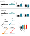
The mGlu7a receptor decreases TRN neurons-mediated tonic and phasic GABAA inhibition in VPM and TRN. (A) Representative current traces of spontaneous mIPSCs recorded from WT and mGlu7AAA TRN neurons in the presence of TTX (1 μM). (B,C) Mean mIPSCs frequency (B) and amplitude (C) in TRN neurons. (D,E,F) Same legend as in (A–C), respectively, in VPM neurons. (G) Example traces of tonic GABAA current recorded in WT and mGlu7AAA TRN neurons upon application of 50 μM gabazine (top traces, black and blue) or gabazine plus ADX71743 (10 μM, bottom traces, orange and red). The baseline shift reflects the inhibition of tonic GABAA current. (H) Average tonic GABAA current amplitude in WT and mGlu7AAA neurons with and without co-application of ADX71743 (10 μM). P < 0.05, ∗∗P < 0.01, ∗∗∗P < 0.001, Mann-and-Whitney test. Error bars indicate SEM.
FIGURE 6
FIGURE 6
The mGlu7 receptor modulates glutamatergic transmission between VPM neurons and cortical layer 4 Fast-Spiking interneurons. (A) Experimental configuration and example of a thalamocortical slice expressing hChR2-mCherry in VPM thalamic neurons and their axons targeting cortical layer 4. LED stimuli (blue light, 488 nm) were directed on layer 4 thalamocortical hChR2-expressing terminals where postsynaptic responses of cortical neurons were recorded. (B,D) Representative current traces recorded from a Fast-Spiking interneuron in response to VPM single (B) or 5 × 10 Hz (D) stimulations, in WT (left) and mGlu7aAAA (right) mice. Blue arrows indicate light stimulations. (C) D,L-AP4-induced EPSC amplitude inhibition (n = 17 for WT, n = 19 for mGlu7aAAA mice). (E) Synaptic depression ratios in WT (left) and mGlu7aAAA mice. Please note the decrease in synaptic depression in the mGlu7AAA mice. (F) Representative current traces showing the increase in EPSC by ADX71743. (G) Summary of the effects of ADX71743 (10 μM) on EPSCs. (H,I) Average EPSC inhibition induced by D,L-AP4 and short-term synaptic depression ratios in excitatory neurons and Regular-Spiking interneurons, respectively. Please note the absence of difference between WT and mGlu7aAAA mice. P < 0.05, Mann-and-Whitney test. Error bars indicate SEM.
FIGURE 7
FIGURE 7
Constitutive activity of mGlu7a receptors in the absence of extracellular glutamate. (A) Normalized IP1 production in HEK cells transfected or not (Mock) with increasing amount of mGlu7a receptor cDNA (n = 3). Note that transfection of mGlu7a receptor in the absence of agonist induces IP1 synthesis. (B) IP1 formation measured in the absence (Base) and presence of the indicated compounds in HEK cells transfected with either wild-type (WT) or a control empty vector (n = 5). P < 0.05, ∗∗∗P < 0.001, Mann-and-Whitney test. Error bars indicate SEM.
FIGURE 8
FIGURE 8
EEG activity induced by treatment with the mGlu7 receptor NAM. (A) Behavioral response of mice WT and mGlu7AAA mice treated with the either vehicle or mGlu7 NAM (100 mg/kg i.p.) represented as percentage of total animals tested (n for each score group is indicated on the histogram bars). The scoring scale is given on the right. (B–F) Example of EEG traces and corresponding short-time Fourier transform representation (frequency over time) are shown for WT mice injected with either the vehicle alone (B) or the mGlu7 NAM (C,D) and for representative mGlu7AAA mice showing spontaneous SWD when treated with the vehicle (E) or with the mGlu7 NAM (F). Traces in (C,D) show, respectively, SWD-like and low frequency activity with recurrent spikes corresponding to K-complexes. The level of wavelet power is represented using the color scale. (G) Latency and duration of the lethargic state observed in WT mice upon ADX71743 administration.
FIGURE 9
FIGURE 9
Summary of findings on functional expression of mGlu7 receptors in the thalamocortical network and alterations brought by the loss of mGlu7 constitutive activity or by the mGlu7aAAA mutation.
None

Similar articles

Cited by

References

    1. Ango F., Prézeau L., Muller T., Tu J. C., Xiao B., Worley P., et al. (2001). Agonist-independent activation of metabotropic glutamate receptors by the intracellular protein Homer. Nature 411 962–965. 10.1038/35082096 - DOI - PubMed
    1. Beenhakker M. P., Huguenard J. R. (2009). Neurons that fire together also conspire together: is normal sleep circuitry hijacked to generate epilepsy? Neuron 62 612–632. 10.1016/j.neuron.2009.05.015 - DOI - PMC - PubMed
    1. Berridge M. J., Taylor C. (1988). “Inositol trisphosphate and calcium signaling,” in Proceedings of the Cold Spring Harbor Symposia on Quantitative Biology (Cold Spring Harbor, NY: Cold Spring Harbor Laboratory; ), 927–933. - PubMed
    1. Bertaso F., Lill Y., Airas J. M., Espeut J., Blahos J., Bockaert J., et al. (2006). MacMARCKS interacts with the metabotropic glutamate receptor type 7 and modulates G protein-mediated constitutive inhibition of calcium channels. J. Neurochem. 99 288–298. 10.1111/j.1471-4159.2006.04121.x - DOI - PubMed
    1. Bertaso F., Zhang C., Scheschonka A., De Bock F., Fontanaud P., Marin P., et al. (2008). PICK1 uncoupling from mGluR7a causes absence-like seizures. Nat. Neurosci. 11 940–948. 10.1038/nn.2142 - DOI - PMC - PubMed

Publication types

MeSH terms