Review
. 2016 May 6:10:35.
doi: 10.3389/fncir.2016.00035. eCollection 2016.

Inhibitory Circuits in Cortical Layer 5

Affiliations
Review

Inhibitory Circuits in Cortical Layer 5

Alexander Naka et al. Front Neural Circuits. .

Abstract

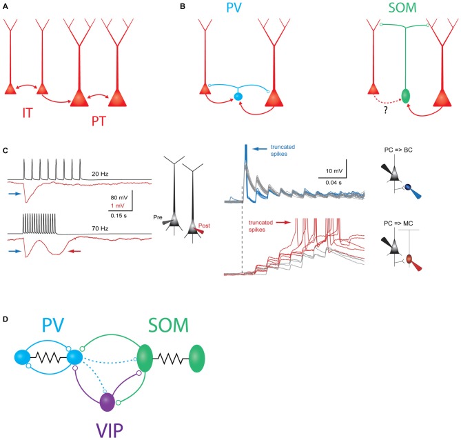
Inhibitory neurons play a fundamental role in cortical computation and behavior. Recent technological advances, such as two photon imaging, targeted in vivo recording, and molecular profiling, have improved our understanding of the function and diversity of cortical interneurons, but for technical reasons most work has been directed towards inhibitory neurons in the superficial cortical layers. Here we review current knowledge specifically on layer 5 (L5) inhibitory microcircuits, which play a critical role in controlling cortical output. We focus on recent work from the well-studied rodent barrel cortex, but also draw on evidence from studies in primary visual cortex and other cortical areas. The diversity of both deep inhibitory neurons and their pyramidal cell targets make this a challenging but essential area of study in cortical computation and sensory processing.

Keywords: barrel cortex; inhibition; inhibitory microcircuits; interneuron; layer 5; neocortex; sensory cortex.


Figures

Figure 1
Figure 1
Schematic overview of major intralaminar circuits in layer 5 (L5). (A) Both the slender-tufted intratelencephalic (IT) cells (left) and thick-tufted pyramidal tract (PT) cells (right) form homotypic excitatory synaptic connections. IT cells additionally connect to PT cells, but PT cells connect to IT cells only very rarely. IT→IT and IT→PT connections both occur at a fairly high rate (Brown and Hestrin, ; Lefort et al., ; Kiritani et al., 2012). PT→PT connectivity occurs less frequently, but is structured into strongly interconnected subnetworks (Song et al., ; Perin et al., 2011). (B) Left: both IT and PT cells (red) excite parvalbumin (PV) cells (blue) and receive perisomatic inhibition from PV cells (Angulo et al., ; Silberberg and Markram, ; Kruglikov and Rudy, 2008). Right: somatostatin (SOM)/Martinotti cells (green) inhibit the distal dendrites of both PT and IT cells. These interneurons receive excitatory input from PT cells, but it is unknown if IT cells also excite them. (C) Experimental evidence for disynaptic inhibitory circuits between L5 pyramidal cells (PCs). Left: example traces showing two types of disynaptic inhibitory responses in a postsynaptic PC (red) driven by spiking in a presynaptic PC (black). Firing the presynaptic cell at 20 Hz (top traces) drives a transient, frequency-independent disynaptic inhibitory response (indicated by the blue arrow) which is likely mediated by activation of a PV/basket cell at the onset of spiking. Firing the same cell at 70 Hz (bottom traces) reveals a second, frequency-dependent form of disynaptic inhibition (indicated by the red arrow) which is likely due to delayed recruitment of a (SOM) Martinotti cell. Right: membrane potential responses of different interneurons to high-frequency stimulation of an L5 PC. Top: (PV) basket cells receive strong excitatory postsynaptic potentials (EPSPs) at the onset of stimulation, which can drive subthreshold (gray traces) or supra-threshold depolarization (blue traces). In either case, the postsynaptic response is initially strong, but then depresses rapidly. Bottom: EPSPs in Martinotti cells are weak and unreliable at the onset of L5 PC firing, but these facilitate and can eventually drive postsynaptic spiking, leading to frequency dependent disynaptic inhibition (FDDI; gray traces, subthreshold responses, red traces- suprathreshold responses). Reproduced with permission from Silberberg and Markram (2007). (D) Schematic of interneuron-to-interneuron connectivity in L5. PV cells (blue) form reciprocal chemical and electrical synapses with other PV cells. SOM cells are electrically but not chemically connected to other SOM cells, and form chemical synapses onto PV cells and vasoactive intestinal peptide (VIP) cells. VIP cells inhibit PV cells and SOM cells. Dashed lines indicate two weaker outputs from PV cells onto SOM cells and VIP cells.
Figure 2
Figure 2
Schematic overview of major translaminar inhibitory circuits impinging on L5 PCs. (A) Inhibition arises from multiple types of interneurons outside of L5. In L1, 5HT3aR/Neurogliaform cells (NGCs, mustard) inhibit L5 PCs via volume release of GABA. In L2/3, SOM/Martinotti cells (green) and VIP cells (purple) target the apical dendrites of L5 PCs, while PV/basket cells (blue) synapse onto the perisomatic compartment via descending axons. In L6, translaminar PV/basket cells inhibit L5 PCs via ascending axons. (B) Excitatory translaminar pathways recruit L5 interneurons. Excitatory cells in L2/3, L4, and L6 can target PV/basket cells. Additionally, L2/3 PCs synapse onto L5 SOM/Martinotti cells.

References

    1. Adesnik H., Bruns W., Taniguchi H., Huang Z. J., Scanziani M. (2012). A neural circuit for spatial summation in visual cortex. Nature 490, 226–231. 10.1038/nature11526 - DOI - PMC - PubMed
    1. Adesnik H., Scanziani M. (2010). Lateral competition for cortical space by layer-specific horizontal circuits. Nature 464, 1155–1160. 10.1038/nature08935 - DOI - PMC - PubMed
    1. Agmon A., Connors B. W. (1989). Repetitive burst-firing neurons in the deep layers of mouse somatosensory cortex. Neurosci. Lett. 99, 137–141. 10.1016/0304-3940(89)90278-4 - DOI - PubMed
    1. Agmon A., Connors B. W. (1992). Correlation between intrinsic firing patterns and thalamocortical synaptic responses of neurons in mouse barrel cortex. J. Neurosci. 12, 319–329. - PMC - PubMed
    1. Andermann M. L., Gilfoy N. B., Goldey G. J., Sachdev R. N. S., Wölfel M., McCormick D. A., et al. . (2013). Chronic cellular imaging of entire cortical columns in awake mice using microprisms. Neuron 80, 900–913. 10.1016/j.neuron.2013.07.052 - DOI - PMC - PubMed

Publication types

LinkOut - more resources