. 2016 May 4:10:83.
doi: 10.3389/fnbeh.2016.00083. eCollection 2016.

Persistent Impact of In utero Irradiation on Mouse Brain Structure and Function Characterized by MR Imaging and Behavioral Analysis

Affiliations

Persistent Impact of In utero Irradiation on Mouse Brain Structure and Function Characterized by MR Imaging and Behavioral Analysis

Tine Verreet et al. Front Behav Neurosci. .

Abstract

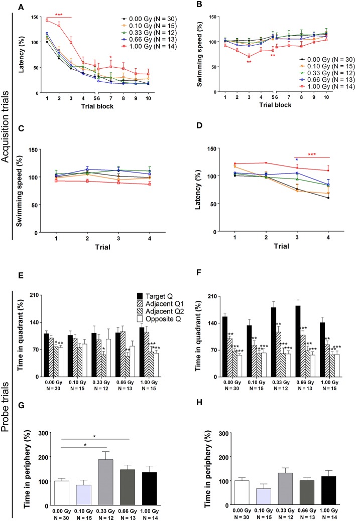
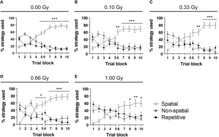
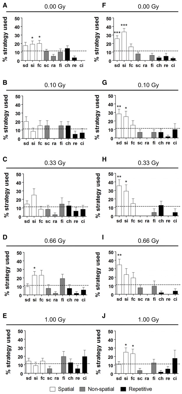
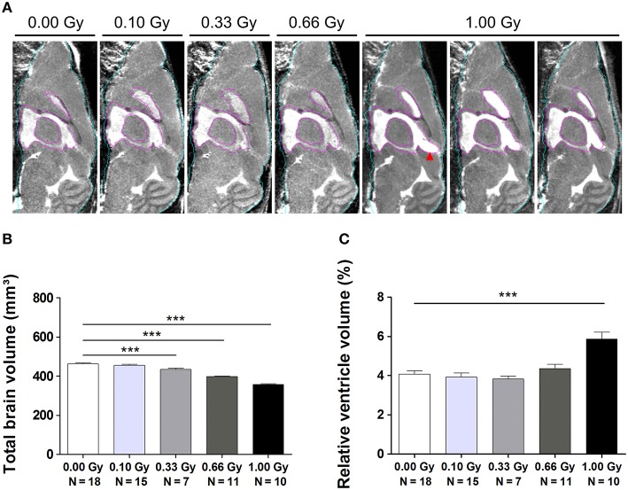
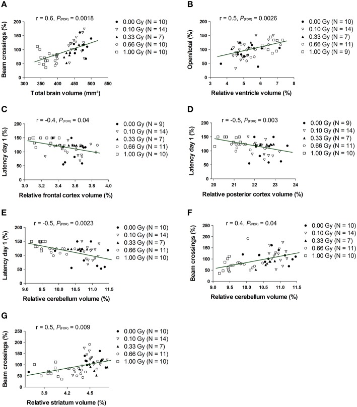
Prenatal irradiation is known to perturb brain development. Epidemiological studies revealed that radiation exposure during weeks 8-15 of pregnancy was associated with an increased occurrence of mental disability and microcephaly. Such neurological deficits were reproduced in animal models, in which rodent behavioral testing is an often used tool to evaluate radiation-induced defective brain functionality. However, up to now, animal studies suggested a threshold dose of around 0.30 Gray (Gy) below which no behavioral alterations can be observed, while human studies hinted at late defects after exposure to doses as low as 0.10 Gy. Here, we acutely irradiated pregnant mice at embryonic day 11 with doses ranging from 0.10 to 1.00 Gy. A thorough investigation of the dose-response relationship of altered brain function and architecture following in utero irradiation was achieved using a behavioral test battery and volumetric 3D T2-weighted magnetic resonance imaging (MRI). We found dose-dependent changes in cage activity, social behavior, anxiety-related exploration, and spatio-cognitive performance. Although behavioral alterations in low-dose exposed animals were mild, we did unveil that both emotionality and higher cognitive abilities were affected in mice exposed to ≥0.10 Gy. Microcephaly was apparent from 0.33 Gy onwards and accompanied by deviations in regional brain volumes as compared to controls. Of note, total brain volume and the relative volume of the ventricles, frontal and posterior cerebral cortex, cerebellum, and striatum were most strongly correlated to altered behavioral parameters. Taken together, we present conclusive evidence for persistent low-dose effects after prenatal irradiation in mice and provide a better understanding of the correlation between their brain size and performance in behavioral tests.

Keywords: MRI; brain development; cognition; microcephaly; radiation exposure; sociability.


Figures

Figure 1
Figure 1
Image registration and labeling. On top, five representative brain MR images are shown. Of all the images from all conditions, we prepared an average study template, which was then registered to the NUS atlas in order to segment the brain into specific brain regions. These brain structures or labels were propagated to the individual images of every animal. The whole brain contour is shown in cyan, the ventricles are delineated in purple, and the frontal and cerebral cortex in red and green, respectively.
Figure 2
Figure 2
Decreased 23-h spontaneous cage activity, with the conservation of the circadian rhythm, is observed in prenatally high-dose exposed mice. (A) Animals irradiated with 1.00 Gy at E11 displayed a decreased activity, as assessed by the total amount of beam crossings during a 23-h period. (B) Examination of the circadian profile of all animals indicated a decline in beam crossings during the dark phase in high-dose exposed mice. Data are presented as mean ± SEM. Horizontal lines with an asterisks indicate significant differences between means of two groups. *P < 0.05, **P < 0.01, ***P < 0.001. The number of animals used is indicated in the graphs (N). E, embryonic day; Gy, Gray.
Figure 3
Figure 3
Mice irradiated in utero with 1.00 Gy spent more time in the open arms of the elevated plus maze as compared to sham-exposed animals. (A) No difference in total arm entries was found. (B) When calculating the ratio between the number of entries in the open arms in relation to the total arm entries, a significant increase was seen for the high-dose exposed mice. These results therefore point toward a decreased inhibition of irradiated mice to enter the open arms. Data are presented as mean ± SEM. Horizontal lines with an asterisks indicate significant differences between means of two groups. *P < 0.05. The number of animals used is indicated in the graphs (N). Gy: Gray.
Figure 4
Figure 4
Prenatally irradiated animals show an increased social behavior, as evidenced by the amount of nose contact in the SPSN task. (A) During the second trial of the SPSN test, all animals exposed to radiation at E11 engaged more time in nose contact with the STR in the wire cage as compared to sham-irradiated animals. (B,C) In the following trial, a second STR, STR2, was placed in the other wire cage and time spent in contact (B), as well as entries in the nose contact zone (C), between STR1 and STR2 were compared. Here, all animals, except for those exposed to 1.00 Gy, showed a significantly increased interest in STR2, indicative for social memory. Data are presented as mean ± SEM. Horizontal lines with an asterisks indicate significant differences between means of two groups. *P < 0.05, **P < 0.01, ***P < 0.001. The number of animals used is indicated in the graphs (N). E, embryonic day; Gy, Gray; SPSN, sociability and preference for social novelty; STR, stranger mouse.
Figure 5
Figure 5
A traditional hidden-platform MWM analysis points toward aberrations in learning and memory in mice irradiated at E11. (A) A 10-day MWM protocol was exploited to evaluate changes in spatial learning and memory between control and in utero irradiated mice. This revealed an increased latency of 1.00-Gy exposed animals to find the hidden platform, which was especially apparent during the first trial days of the test. (B) During these trials, the high-dose exposed mice displayed a lower swimming speed. (C,D) More careful examination of the first trial day showed that significant changes in escape latency were already visible in 0.66- and 1.00-Gy irradiated animals (C), while the decreased swimming velocity in high-dose irradiated animals was not that evident yet (D). (E,F) The probe trials performed on day 6 (E) and 11 (F) did not indicate a pronounced effect of irradiation on quadrant preference. Asterisks indicate significant differences in time spent in the respective quadrants in respect to the target quadrant for each experimental group. (G,H) Notably, mice exposed to a moderate irradiation dose showed an elevated time in swimming in the periphery during the probe trial at day 6 (G), but this difference disappeared in the day 11 probe trial (H). Data are presented as mean ± SEM. *P < 0.05, **P < 0.01, ***P < 0.001. The number of animals used is indicated in the graphs (N). E, embryonic day; Gy, Gray; MWM, Morris water maze; Q, quadrant.
Figure 6
Figure 6
Swim strategy analysis discloses differences in higher cognitive functioning between dose groups. (A–E) We used the analysis of swim strategies in the MWM acquisition trials to uncover subtle dissimilarities in higher spatio-cognitive performance in control animals (A) vs. mice in utero exposed to 0.10 (B), 0.33 (C), 0.66 (D), or 1.00 Gy (E). The exclusive use of the spatial strategy in sham-exposed mice from day 5 onwards was not found in the other dose groups. Indeed, mice irradiated with 0.10 Gy only showed this predominant spatial strategy use from training day 6 on and 0.33-Gy irradiated mice from day 7 on. The most pronounced differences in this analysis were observed in the animals exposed to the highest dose of 1.00 Gy (exclusive use of spatial strategy from day 8 on). Conflicting to this dose-response relationship, the 0.66-Gy exposed animals already significantly used their spatial strategy as compared to the other two strategy classes from training day 4 onwards. Data are presented as mean ± SEM. Horizontal lines with an asterisks indicate significant differences between the spatial vs. non-spatial and repetitive strategies for all the trial blocks underneath. *P < 0.05, **P < 0.01, ***P < 0.001. Gy, Gray; MWM, Morris water maze.
Figure 7
Figure 7
Irradiated mice have differential search strategy profiles halfway and at the end of MWM training. (A–J) Low-dose effects on spatial memory were confirmed when detailed strategy profiles were examined at day 5 (A–E) and 10 (F–J) of the MWM acquisition training. Whereas controls had a significant preference for spatial strategies on training day 5, this was not observed for the other doses, except for the 0.66-Gy irradiated animals that preferentially used the “spatial indirect” strategy. At training day 10, the two highest dose groups (0.66 and 1.00 Gy) still showed alterations in their preference profiles as compared to the sham-exposed condition, with only a significant use of “spatial direct” for the 0.66-Gy group and a significant use of both the “spatial indirect” and “focal correct” strategies in the 1.00-Gy exposed mice. Data are presented as mean ± SEM. *P < 0.05, **P < 0.01, ***P < 0.001. ch, chaining; ci, circling; fc, focal correct; fi, focal incorrect; Gy, Gray; MWM, Morris water maze; pe, peripheral looping; ra, random; sc, scanning; sd, spatial direct; si, spatial indirect.
Figure 8
Figure 8
MRI unveils microcephaly and enlarged ventricles in in utero irradiated mice. (A) MIPs of representative MR images for all dose groups. The quality of atlas-based label propagation was visually verified using these MIPs. Whole-brain outline is shown in cyan, the delineation of the lateral and third ventricles is presented in purple. Animals irradiated with 1.00 Gy typically demonstrated a swelling of the dorsal part of the third ventricle (red arrowhead). (B) Total brain volume decreased drastically depending on the radiation dose, with a significant decline already observed in 0.33-Gy exposed mice. (C) Ventricles of animals prenatally exposed to 1.00 Gy were increased in size in relation to the total brain volume. Data are presented as mean ± SEM. Horizontal lines with an asterisks indicate significant differences between means of two groups. ***P < 0.001. The number of animals used is indicated in the graphs (N). Gy: Gray. MIP: maximum intensity projection. MRI: magnetic resonance imaging.
Figure 9
Figure 9
The whole-brain volume, along with the normalized volumes of the ventricles, frontal and posterior cerebral cortex, cerebellum and striatum, correlate significantly with altered behavioral outcome. Statistically significant correlations were found between the total brain volume and number of beam crossings in the cage activity (A) and between the normalized ventricle volume and the number of open/total (B) and open/closed (not shown) arm entries in the elevated plus maze. Furthermore, we discovered correlative relationships between the normalized frontal cortex volume and MWM latency (C), between the normalized posterior cortical volume and MWM latency (D), between the normalized cerebellar volume and MWM latency (E), between the normalized cerebellar volume and cage activity beam crossings (F) and between the normalized striatal volume and cage activity beam crossing (G). Correlation coefficients (r) and P values are indicated for each correlation. The number of animals used is indicated in the legend (N). FDR, False discovery rate; Gy, Gray; MWM, Morris water maze; SPSN, Sociability and preference for social novelty.

Similar articles

Cited by

References

    1. Aldridge K., Wang L., Harms M. P., Moffitt A. J., Cole K. K., Csernansky J. G., et al. . (2012). A longitudinal analysis of regional brain volumes in macaques exposed to X-irradiation in early gestation. PLoS ONE 7:e43109. 10.1371/journal.pone.0043109 - DOI - PMC - PubMed
    1. Atwood T., Payne V. S., Zhao W., Brown W. R., Wheeler K. T., Zhu J. M., et al. . (2007a). Quantitative magnetic resonance spectroscopy reveals a potential relationship between radiation-induced changes in rat brain metabolites and cognitive impairment. Radiat. Res. 168, 574–581. 10.1667/RR0735.1 - DOI - PubMed
    1. Atwood T., Robbins M. E., Zhu J. M. (2007b). Quantitative in vivo proton MR spectroscopic evaluation of the irradiated rat brain. J. Magn. Reson. Imaging 26, 1590–1595. 10.1002/jmri.21095 - DOI - PubMed
    1. Bai J., Trinh T. L., Chuang K. H., Qiu A. (2012). Atlas-based automatic mouse brain image segmentation revisited: model complexity vs. image registration. Magn. Reson. Imaging 30, 789–798. 10.1016/j.mri.2012.02.010 - DOI - PubMed
    1. Baskar R., Devi P. U. (2000). Influence of gestational age to low-level gamma irradiation on postnatal behavior in mice. Neurotoxicol. Teratol. 22, 593–602. 10.1016/S0892-0362(00)00076-3 - DOI - PubMed

LinkOut - more resources