

The Physiology of Bone Pain. How Much Do We Really Know?
- PMID: 27199772
- PMCID: PMC4844598
- DOI: 10.3389/fphys.2016.00157
The Physiology of Bone Pain. How Much Do We Really Know?
Abstract
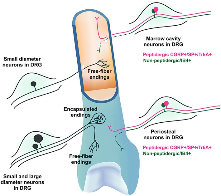
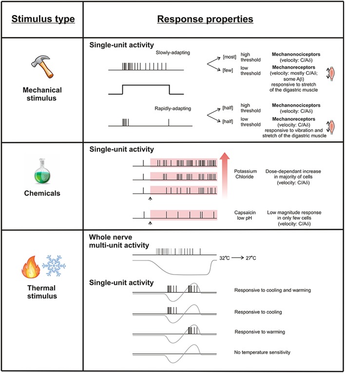
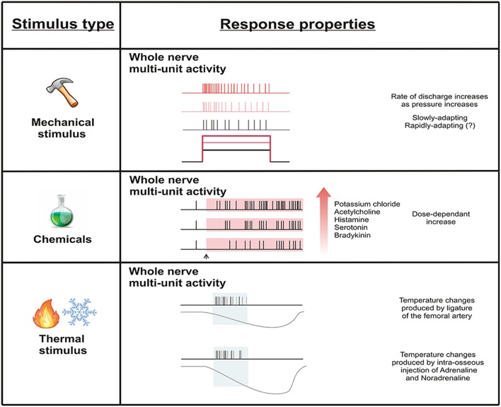
Pain is associated with most bony pathologies. Clinical and experimental observations suggest that bone pain can be derived from noxious stimulation of the periosteum or bone marrow. Sensory neurons are known to innervate the periosteum and marrow cavity, and most of these have a morphology and molecular phenotype consistent with a role in nociception. However, little is known about the physiology of these neurons, and therefore information about mechanisms that generate and maintain bone pain is lacking. The periosteum has received greater attention relative to the bone marrow, reflecting the easier access of the periosteum for experimental assessment. With the electrophysiological preparations used, investigators have been able to record from single periosteal units in isolation, and there is a lot of information available about how they respond to different stimuli, including those that are noxious. In contrast, preparations used to study sensory neurons that innervate the bone marrow have been limited to recording multi-unit activity in whole nerves, and whilst they clearly report responses to noxious stimulation, it is not possible to define responses for single sensory neurons that innervate the bone marrow. There is only limited evidence that peripheral sensory neurons that innervate bone can be sensitized or that they can be activated by multiple stimulus types, and at present this only exists in part for periosteal units. In the central nervous system, it is clear that spinal dorsal horn neurons can be activated by noxious stimuli applied to bone. Some can be sensitized under pathological conditions and may contribute in part to secondary or referred pain associated with bony pathology. Activity related to stimulation of sensory nerves that innervate bone has also been reported in neurons of the spinoparabrachial pathway and the somatosensory cortices, both known for roles in coding information about pain. Whilst these provide some clues as to the way information about bone pain is centrally coded, they need to be expanded to further our understanding of other central territories involved. There is a lot more to learn about the physiology of peripheral sensory neurons that innervate bone and their central projections.
Keywords: bone; bone marrow; bone pain; electrophysiology; nociception; pain; periosteum.
Figures
References
-
- Arnoldi C. C. (1990). Intraosseous engorgement-pain syndromes. The pathomechanism of pain, in Bone Circulation and Bone Necrosis, eds Arlet J., Mazières B. (Berlin; Heidelberg: Springer; ), 253–259. 10.1007/978-3-642-73644-5_52 - DOI
Publication types
LinkOut - more resources
Full Text Sources
Other Literature Sources