. 2016 May 19;62(4):572-85.
doi: 10.1016/j.molcel.2016.04.016.

Recognition of Lys48-Linked Di-ubiquitin and Deubiquitinating Activities of the SARS Coronavirus Papain-like Protease

Affiliations

Recognition of Lys48-Linked Di-ubiquitin and Deubiquitinating Activities of the SARS Coronavirus Papain-like Protease

Miklós Békés et al. Mol Cell. .

Abstract

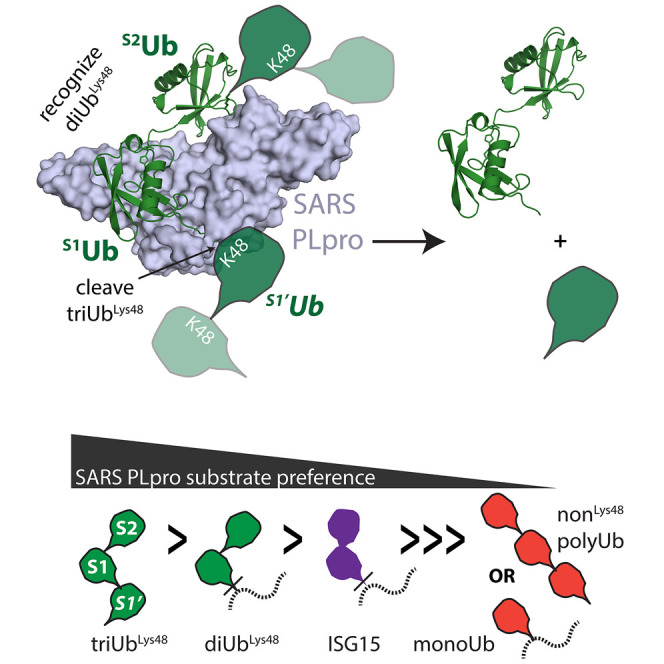
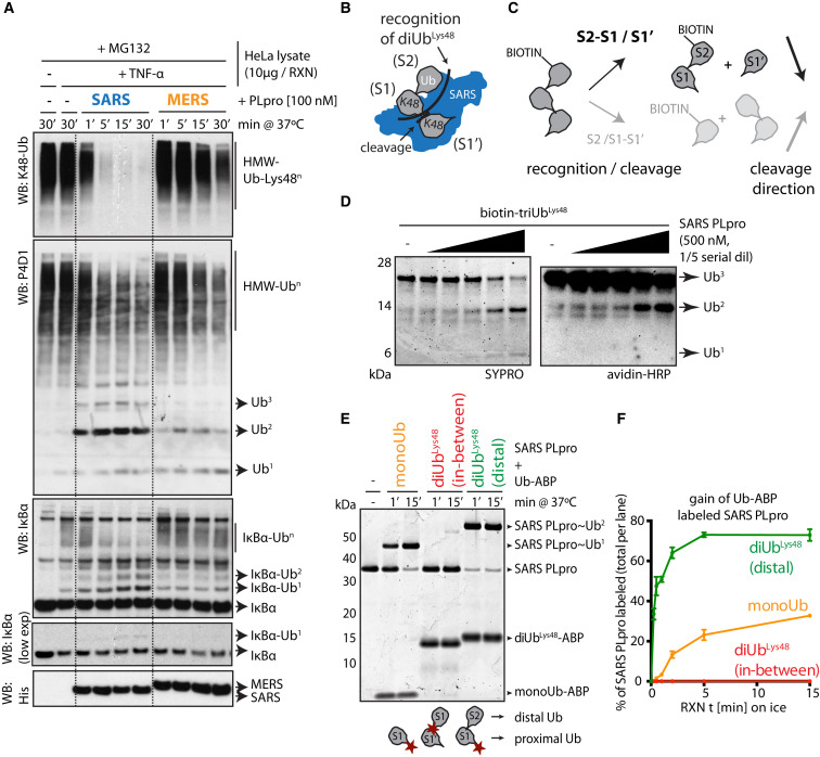
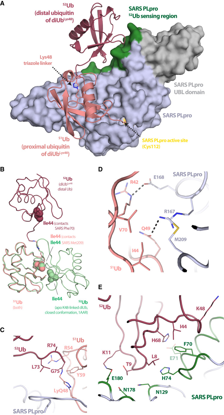
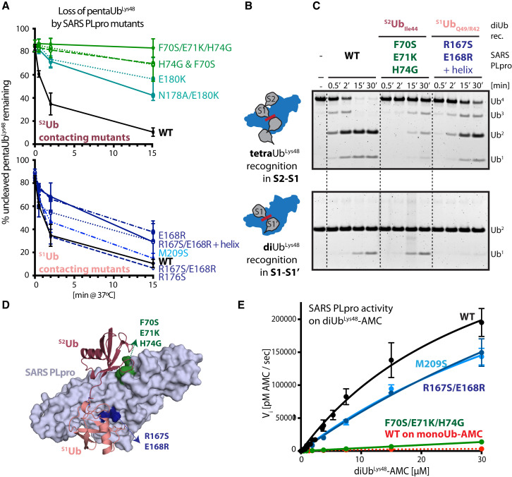
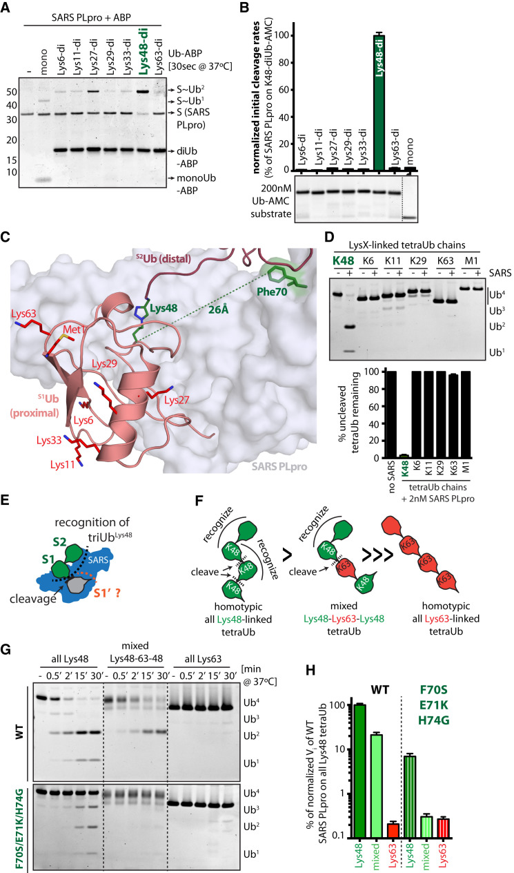
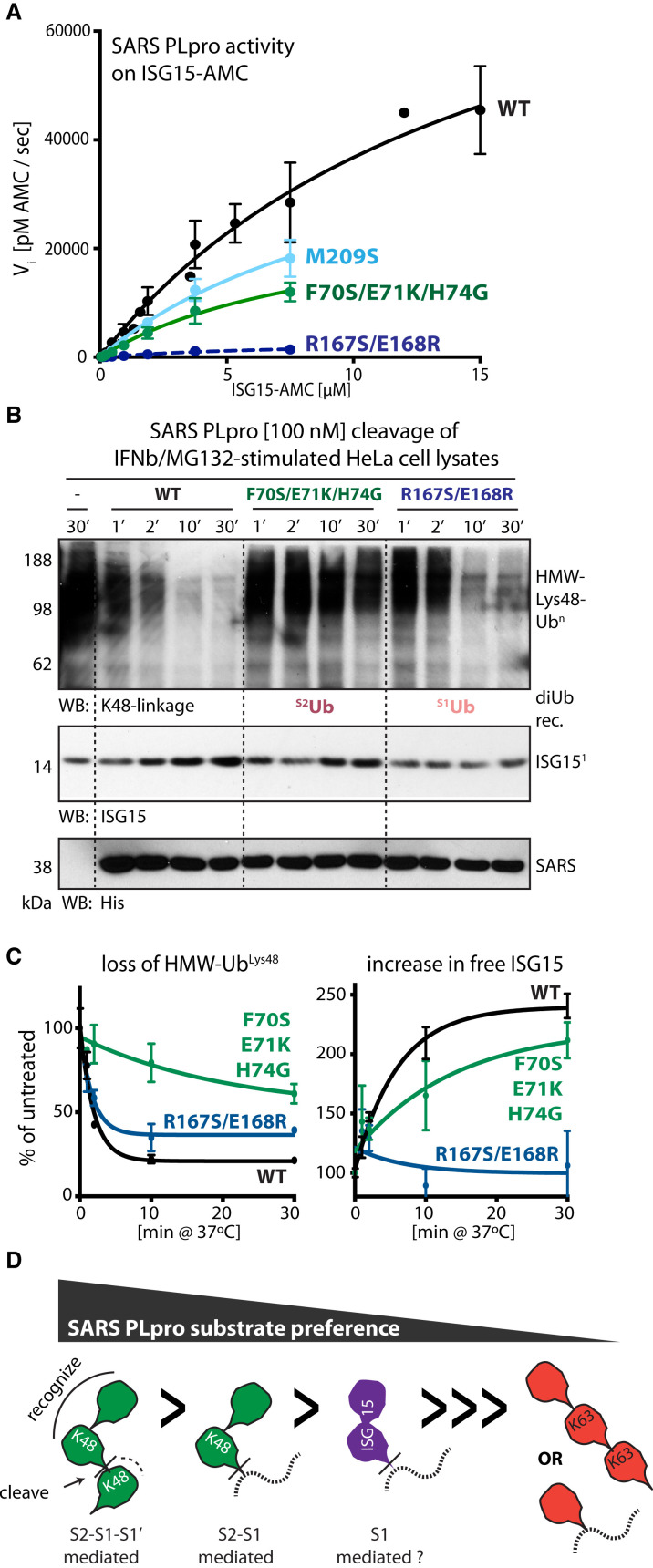
Deubiquitinating enzymes (DUBs) recognize and cleave linkage-specific polyubiquitin (polyUb) chains, but mechanisms underlying specificity remain elusive in many cases. The severe acute respiratory syndrome (SARS) coronavirus papain-like protease (PLpro) is a DUB that cleaves ISG15, a two-domain Ub-like protein, and Lys48-linked polyUb chains, releasing diUb(Lys48) products. To elucidate this specificity, we report the 2.85 Å crystal structure of SARS PLpro bound to a diUb(Lys48) activity-based probe. SARS PLpro binds diUb(Lys48) in an extended conformation via two contact sites, S1 and S2, which are proximal and distal to the active site, respectively. We show that specificity for polyUb(Lys48) chains is predicated on contacts in the S2 site and enhanced by an S1-S1' preference for a Lys48 linkage across the active site. In contrast, ISG15 specificity is dominated by contacts in the S1 site. Determinants revealed for polyUb(Lys48) specificity should prove useful in understanding PLpro deubiquitinating activities in coronavirus infections.


Figures

None
Graphical abstract
Figure 1
Figure 1
Distal diUbLys48 ABP Labels SARS PLpro (A) Ub-conjugate cleavage in TNF-α-treated HeLa cell lysates by SARS and MERS PLpro. Dotted lines added for clarity. (B and C) Schematics of (B) Ub chain recognition by SARS PLpro and (C) recognition and cleavage of biotin-tagged triUbLys48. (D) Cleavage of biotin-triUbLys48 by SARS PLpro. Cleavage intermediates detected by avidin-HRP reveal biotin on the diUb product. (E) Qualitative labeling of SARS PLpro by Ub-ABPs (cartoons at bottom with red stars indicate warhead positions). (F) Quantitative labeling of SARS PLpro by Ub-ABPs indicating percent of SARS PLpro labeled as derived from gels in Figure S2C. Error bars represent ±SEM. See also Figures S1 and S2.
Figure 2
Figure 2
Crystal Structure of SARS PLpro Bound to a diUbLys48-ABP (A) Cartoon and surface representations of SARS PLpro covalently bound to diUbLys48-ABP. SARS PLpro shown in surface representation with the USP-family DUB domain colored blue-white, the N-terminal Ubl domain in gray, the S2Ub binding motif in green, and the active site cysteine (C112) in yellow. Cartoon representation of Lys48-linked diUb with proximal Ub (S1Ub) in salmon and distal Ub (S2Ub) in raspberry. (B–E) Cartoon representation of interaction surfaces between SARS PLpro and diUbLys48; color as in (A). (B) Structure-based alignment of SARS-PLpro-diUbLys48 and a closed conformation of diUbLys48 (PDB 1AAR). Ub Ile44 (hydrophobic patch) that contact SARS PLpro shown as spheres. (C) View of the covalent triazole linkage between S1Ub-Lys48 and S2Ub-Gly75 indicating minimal contacts to SARS PLpro. (D) View of contacts between the SARS PLpro palm domain (M209 and Arg167/Asp168) and the S1Ub, highlighting both hydrophobic and polar interactions (S1Ub-Ile44 and -Gln49/Arg42), respectively. (E) View of contacts between the SARS PLpro S2Ub binding motif (Phe70, Glu71, and His74) and the S2Ub-Ile44 hydrophobic patch (S2Ub-Ile44, -Leu8, and -His68). See also Figure S3.
Figure 3
Figure 3
Contribution of SARS PLpro S1Ub and S2Ub Sites to polyUbLys48 Cleavage (A) Ub chain cleavage activities of SARS PLpro WT (black) and its mutants (S2Ub, top, green; S1Ub, bottom, blue) on pentaUbLys48. Representative gels used to derive graphs shown in Figure S3B. Error bars represent ±SEM. (B) Schematics of tetraUbLys48 and diUbLys48 recognition by SARS PLpro. (C) Gel-based cleavage assays of SARS PLpro WT and mutants on tetraUbLys48 (top) and diUbLys48 (bottom) indicating differential effects of diUb Lys48 and tetraUb Lys48 cleaving activities for S1Ub and S2Ub mutants of SARS PLpro. Additional mutants analyzed in Figure S4C. (D) Cartoon and surface representation of SARS-PLpro∼diUbLys48 indicating the location of the S1Ub and S2Ub mutants. (E) Michaelis-Menten kinetics of WT (black) and selected SARS PLpro mutants (M209S, hydrophobic S1 mutant, light blue; R167S/E168R, polar S1 mutant, dark blue; F70S/E71K/H74G, S2 mutant, green) on diUbLys48-AMC substrates and comparison to SARS PLpro WT on monoUb-AMC (in red). Extracted kinetic parameters (kcat and KM) are in Table 2. See also Figure S4.
Figure 4
Figure 4
SARS PLpro Activity Is Restricted to Lys48-Linked Ub Chains with Specificity Dominated by S2-S1 Interactions (A and B) S2-S1 linkage specificity of SARS PLpro probed by (A) SDS-PAGE analysis of cross-linking to linkage-specific diUb-ABPs (SYPRO-stained) and (B) release of AMC using diUb-AMC fluorogenic substrates with initial linear cleavage rates (Vi) plotted as percent of diUbLys48-AMC cleavage rate by WT SARS PLpro as derived from curves in Figure S4B. (C) View of SARS-PLpro∼diUbLys48-ABP highlighting the location of S1Ub-Lys48 (dark green, sticks) and its proximity to Phe70 (green) in the SARS PLpro S2Ub binding site. Other chain-forming S1Ub residues (Lys-6, -11, -27, -29, -33, and -63 and Met1) shown as sticks (red). (D) Linkage specificity of SARS PLpro assayed using homotypic tetraUb chains. Representative gel shown and bar graph indicating ±SEM from duplicate experiments. (E) Schematics of triUbLys48 chain recognition by SARS PLpro via S2-S1 (green) and S1′ (gray). (F) Cartoon of tetraUb chains, indicating SARS PLpro preferred sites of recognition (curved line) and cleavage (dotted line). (G) Time-course for cleavage of tetraUb chains by SARS PLpro and its S2 mutant. (H) Quantification of Vi as determined from duplicate experiments in Figure 4G. Error bars represent ±SEM. See also Figure S5.
Figure 5
Figure 5
Recognition of DiUbLys48 and ISG15 by SARS PLpro Appears Distinct (A) Michaelis-Menten kinetics of WT (black) and selected SARS PLpro mutants (M209S, S1 mutant, light blue; R167S/E168R, S1 mutant, dark blue; F70S/E71K/H74G, S2 mutant, green) using ISG15-AMC. Extracted kinetic parameters (kcat and KM) in Table 2. (B) Cleavage of HMW-UbLys48 (top, WB anti-K48) and ISG15-conjugates (bottom, WB ISG15) in lysates prepared from IFNβ/MG132-treated cells by SARS PLpro WT and S2 and S1 mutants. (C) Quantification of loss of HMW-UbLys48 (left) and appearance of free ISG15 (right) from duplicate experiments shown in Figure 5B. Error bars represent ±SEM. (D) Schematic representation of SARS PLpro substrate specificity. Dashed lines can indicate the -AMC substrate, a non-Lys48-linked Ub unit, or a protein substrate. See also Figure S6.

References

    1. Báez-Santos Y.M., Mielech A.M., Deng X., Baker S., Mesecar A.D. Catalytic function and substrate specificity of the papain-like protease domain of nsp3 from the Middle East respiratory syndrome coronavirus. J. Virol. 2014;88:12511–12527. - PMC - PubMed
    1. Bailey-Elkin B.A., Knaap R.C., Johnson G.G., Dalebout T.J., Ninaber D.K., van Kasteren P.B., Bredenbeek P.J., Snijder E.J., Kikkert M., Mark B.L. Crystal structure of the Middle East respiratory syndrome coronavirus (MERS-CoV) papain-like protease bound to ubiquitin facilitates targeted disruption of deubiquitinating activity to demonstrate its role in innate immune suppression. J. Biol. Chem. 2014;289:34667–34682. - PMC - PubMed
    1. Bailey-Elkin B.A., van Kasteren P.B., Snijder E.J., Kikkert M., Mark B.L. Viral OTU deubiquitinases: a structural and functional comparison. PLoS Pathog. 2014;10:e1003894. - PMC - PubMed
    1. Békés M., Rut W., Kasperkiewicz P., Mulder M.P., Ovaa H., Drag M., Lima C.D., Huang T.T. SARS hCoV papain-like protease is a unique Lys48 linkage-specific di-distributive deubiquitinating enzyme. Biochem. J. 2015;468:215–226. - PMC - PubMed
    1. Berger A., Schechter I. Mapping the active site of papain with the aid of peptide substrates and inhibitors. Philos. Trans. R. Soc. Lond. B Biol. Sci. 1970;257:249–264. - PubMed

MeSH terms

LinkOut - more resources